Musculoskeletal Ultrasound: The Complete Guide, Joint by Joint
The principles that apply to any joint (compartments, laterality, measurements, dynamic exam, Doppler and the limits of the method) and the roadmap for the shoulder, hand and wrist, knee, ankle and foot, soft tissues and muscles.
 LaudárioUltrasound reports in practice
Musculoskeletal ultrasound has an advantage no other imaging method offers: examining the structure at the exact site of pain, in motion and comparing with the other side at the same moment. It also has a weakness: it is the exam in which generic reports are most common, with "tendinopathy" and no mention of which tendon, at what point and with what measurement.
This guide gathers the principles that apply to any joint and points, region by region, to the article that goes deeper into each one. Use it as an exam roadmap and as an index of what we have already written on musculoskeletal imaging.
Principles that apply to all joints
- Examine by compartments, always in the same order. Anterior, medial, lateral and posterior, or dorsal and volar. A fixed order prevents forgetting structures and makes clear in the report what was evaluated.
- Laterality in every sentence. Right or left and, for the fingers, which finger. It seems obvious, and it is the most common mistake.
- Compare with the contralateral side when the finding is subtle. Tendon and fascia thickening are relative.
- Measure. Thickness of tendon, fascia and pulley, nerve area, tear size and gap between stumps. The number is what allows comparison at follow-up.
- Use the dynamic exam when the complaint is catching, subluxation or impingement. It shows what the static exam does not.
- Use Doppler for neovascularization in tendinopathy, active synovitis, and to avoid mistaking an aneurysm for a cyst.
- State what ultrasound does not evaluate well in that region (medial meniscus, cruciate ligaments, deep cartilage, bone lesions) and recommend MRI when the clinical question depends on it.
Shoulder
This is the most requested musculoskeletal exam. The focus is the rotator cuff: supraspinatus, infraspinatus, subscapularis, teres minor and the long head of the biceps, as well as the subacromial-subdeltoid bursa and the impingement maneuvers. In a tear, the report states whether it is partial (and on which surface) or complete, the measurements in both axes and the retraction. See the shoulder report.
Hand, wrist and peripheral nerves
The most common complaints are De Quervain (with the intracompartmental septum that changes the injection and the surgery), trigger finger (the A1 pulley), ganglion cyst (with the origin and pedicle) and carpal tunnel. See hand and wrist.
In compressive neuropathies, the nerve cross-sectional area, measured at the right point, is the number the report needs to include. See peripheral nerves and cross-sectional area.
Knee
Ultrasound evaluates the extensor apparatus (quadriceps and patellar tendons), the collateral ligaments, the bursae, joint effusion and synovitis well, and the interior of the joint poorly. The report needs to make this limit clear. See the knee report.
In the popliteal fossa, the Baker cyst is confirmed by the neck between the semimembranosus and the medial gastrocnemius. When ruptured, it mimics thrombosis, and the deep veins need to be evaluated in the same exam. See Baker cyst.
Ankle and foot
The Achilles tendon is described as insertional or noninsertional tendinopathy, because treatment differs; a tear is reported with the gap between the stumps. The exam is completed by the peroneal tendons (with the dynamic maneuver for subluxation and the search for a longitudinal split), the posterior tibial tendon, the ligaments of the sprain, the plantar fascia thickness and the intermetatarsal space for Morton neuroma. See ankle and foot.
Soft tissues and wall
Subcutaneous nodules (lipomas, epidermal cysts, hematomas, vascular lesions) and abdominal and inguinal wall hernias are frequent exams, and follow the same logic: location by anatomic plane, measurements, content, vascularity and maneuvers. See soft tissues and abdominal wall hernias.
In any region, a solid lesion without typical benign features, especially if deep to the fascia, larger than 5 cm or growing, deserves investigation with MRI.
Muscles
In sports muscle injuries, the report describes the muscle, the location (myotendinous junction, belly, insertion), the extent of fiber disruption, the hematoma with measurements and, at follow-up, healing. The grading system used by the service should be cited, so that the sports physician can compare between exams.
The mistakes that appear most in the musculoskeletal report
- Finding without laterality or without the finger.
- "Tendinopathy" without the tendon, the point and the thickness.
- Tear without measurements, without retraction or without the gap between stumps.
- Complaint of catching without a dynamic exam.
- Mass in the popliteal fossa without Doppler.
- Not stating what ultrasound does not evaluate in that region.
How Laudário helps
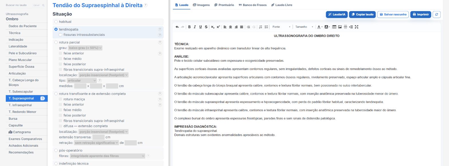
Laudário has dedicated templates for the shoulder, elbow, wrist, hand, hip, knee, ankle and foot, as well as peripheral nerves, muscle, soft tissues and the rheumatologic wrist and hand. In practice:
- Compartments organized in tabs, with laterality.
- Ready-made phrases for tendinopathy, tear, tenosynovitis, bursitis and synovitis, with the measurement fields in the right place.
- Recommendations in their own tab, separate from the findings.
Try it for 15 days free, no credit card.
The ultrasound reporting platform built by sonologists. We write about reports, classifications and standardization from the real routine of the practice. Who writes and how.
Educational content for healthcare professionals. It does not replace clinical judgment or the guidelines adopted by your service. Editorial policy.