Shoulder ultrasound: how to structure the musculoskeletal report with clarity and standardization
How to structure the shoulder ultrasound report with clarity: rotator cuff, biceps, bursa, acromioclavicular joint and dynamic maneuvers.
 LaudárioUltrasound reports in practice
This article is part of the guide Musculoskeletal Ultrasound: The Complete Guide, Joint by Joint.
Shoulder ultrasound is today one of the most frequently requested musculoskeletal exams in outpatient practice, especially for investigating chronic pain, suspected rotator cuff injury and postoperative follow-up. Even so, it is also one of the reports with the greatest variability between professionals: some descriptions cover only the supraspinatus, others forget to measure the bursa, and few systematically comment on dynamic maneuvers. When the report is incomplete, the orthopedist loses relevant clinical information and the patient ends up repeating exams. In this article, we lay out a practical structure for the shoulder ultrasound report, focused on the rotator cuff, long head of the biceps, subacromial-subdeltoid bursa, acromioclavicular joint and dynamic evaluation.
Why does the shoulder report require such a rigorous structure?
The shoulder brings together, in a small space, several structures that must be evaluated individually: four rotator cuff tendons, the long head of the biceps, two bursae, the acromioclavicular joint and the bony interface of the humeral head and tuberosities. Without a fixed sequence, three problems appear frequently:
- Incomplete evaluation of the rotator cuff: the report describes the supraspinatus in detail but treats the infraspinatus, teres minor and subscapularis generically, losing information about posterior and anterior lesions.
- Imprecise description of tendon lesions: “tendinopathy” without stating which tendon, in which portion (insertion, myotendinous junction or muscle belly), whether there is a partial articular-sided, bursal-sided or intrasubstance tear, and the extent of the lesion.
- Absence of dynamic maneuvers: not commenting on the search for subacromial impingement, the behavior of the long head of the biceps tendon in internal and external rotation or the stability of the acromioclavicular joint under stress takes away part of the exam's value compared with MRI.
Structuring the shoulder report is, above all, making sure that all of the functional anatomy is always covered: biceps, subscapularis, supraspinatus, infraspinatus and teres minor, bursa, acromioclavicular joint and dynamic maneuvers, closing with a prioritized and clinically useful impression.
Recommended structure for the shoulder ultrasound report
A good structure follows the classic shoulder scanning sequence, starting with the anterior portion and progressing to the lateral and posterior. This order helps with typing while scanning and reduces the risk of skipping structures. We suggest the following blocks:
1. Identification and technique
State the type of exam (shoulder ultrasound, with dynamic evaluation), side examined, equipment and transducer when relevant, patient positioning and any technical limitations: significant pain that restricted maneuvers, body habitus, marked edema, dressings or fixation hardware. This block protects the professional and tells the orthopedist what could actually be evaluated.
2. Long head of the biceps tendon
Evaluate the tendon in both transverse and longitudinal planes, describing its position in the bicipital groove, thickness, echotexture, echogenicity and the presence of fluid in the sheath. State whether there are signs of tendinopathy, partial tear, medial subluxation or complete dislocation, and describe the status of the biceps pulley when assessable. A small isolated sliver of fluid in the biceps sheath should be interpreted with caution and correlated with a joint effusion.
3. Subscapularis tendon
Examine the subscapularis in external rotation, describing fiber continuity, echotexture, the presence of tears (partial articular-sided, bursal-sided or full-thickness), avulsion of the insertion on the lesser tuberosity, calcifications and associated bursitis. Always state whether or not there is medial migration of the biceps tendon, which is an important indirect sign of subscapularis injury.
4. Supraspinatus tendon
The supraspinatus is the most studied tendon and the most frequently injured. Describe it in as much detail as possible:
- Fiber continuity: intact tendon, with focal thinning, with a partial articular-sided tear, partial bursal-sided tear, intrasubstance tear or full-thickness tear.
- Location of the lesion: insertion on the greater tuberosity, critical zone, myotendinous junction or muscle belly.
- Tear dimensions: extent in the anteroposterior direction and depth in the craniocaudal direction, with measurements in millimeters in both planes.
- Tendon retraction: distance between the tendon stump and the greater tuberosity in complete tears, essential information for surgical planning.
- Appearance of the muscle belly: preserved trophism or atrophy/fatty infiltration, findings that weigh on the surgical indication.
- Intratendinous calcifications: location, dimensions and pattern (dense, with acoustic shadowing, or in the resorptive phase).
Standardized descriptions reduce ambiguity and allow comparison between successive exams of the same patient, especially in follow-up of small tears and in postoperative patients.
5. Infraspinatus and teres minor tendons
Examine both in internal rotation, describing continuity, echotexture, presence of tears and calcifications separately. The teres minor is usually cited generically but deserves its own description, especially in patients with persistent posterior pain or under investigation for Parsonage-Turner syndrome.
6. Subacromial-subdeltoid bursa
State the bursal thickness on a longitudinal view over the supraspinatus, the presence and estimated amount of fluid, the echogenicity of the contents (anechoic, with debris, synovial thickening), septations and signs of bursitis. Reactive bursitis associated with cuff lesions is a relevant finding and should appear clearly in the report, with laterality and anatomical relationship.
7. Acromioclavicular joint
Evaluate the acromioclavicular joint in the coronal plane, describing the regularity of the bony contours, capsular thickness, presence of osteophytes, capsular thickening, joint effusion, synovial cysts and instability on a stress maneuver, when indicated. In patients with shoulder pain on elevation above 90 degrees, this block carries considerable clinical weight.
8. Glenohumeral joint, bony interface and adjacent musculature
Briefly comment on the presence of glenohumeral joint effusion accessible to ultrasound (posterior recess), the regularity of the cortex of the humeral head and tuberosities, the presence of irregularities suggesting a Hill-Sachs lesion in patients with a history of dislocation, and the trophism of the supraspinatus and infraspinatus muscles in their respective fossae.
9. Dynamic evaluation
Explicitly describe the maneuvers performed and their findings: search for subacromial impingement on abduction, behavior of the long head of the biceps tendon in internal and external rotation, stability of the acromioclavicular joint under stress, and evaluation of the subscapularis in external rotation. Dynamic evaluation is a real advantage of ultrasound over MRI and must be recorded in the report.
10. Diagnostic impression and suggested management
The conclusion should be objective, prioritized and geared toward the clinical decision. Begin with the findings of greatest consequence (tendon tears, symptomatic calcifications, large bursitis and signs of impingement) and finish with minor findings and suggested correlations. When applicable, indicate whether there is a benefit in correlation with MRI (for example, in suspected Bankart or SLAP lesions), preserving the orthopedist's role in the final decision.
Common errors in the shoulder report
Even with a defined structure, some recurring errors undermine the quality of the shoulder report:
- Focusing only on the supraspinatus: treating the other cuff tendons generically, without specific description of the subscapularis, infraspinatus and teres minor.
- Not measuring tears: describing a “partial tear” without stating extent and depth prevents the orthopedist from comparing successive exams and planning surgical management.
- Forgetting tendon retraction: in complete tears, omitting the distance between the stump and the greater tuberosity removes from the report a critical piece of data for the surgical indication.
- Treating the bursa vaguely: “bursitis” without thickness, without volume and without characteristics of the contents leaves the finding of little practical use.
- Omitting dynamic maneuvers: static shoulder ultrasound differs little from MRI; the dynamic exam is its greatest advantage and must be described.
- Conclusion disconnected from the description: a detailed descriptive text and a generic impression such as “signs of tendinopathy” create reading noise and impair clinical correlation.
How Laudário helps structure shoulder reports
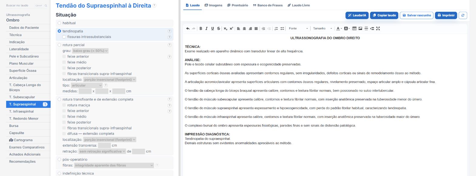
Laudário was designed for professionals who want to report quickly without giving up consistency between exams. For shoulder ultrasound, and for the main musculoskeletal protocols, the system offers:
- Ready-made, structured templates for shoulder, knee, ankle, wrist, elbow and hip, organized in the classic scanning order, to speed up typing without omissions.
- Standardized, customizable phrases for describing each rotator cuff tendon, biceps, bursa, acromioclavicular joint and dynamic maneuvers, keeping terminology consistent between reports of the same patient.
- Specific fields for measurements of tears, tendon retraction, bursal thickness and calcifications, which are available in the body of the report in an organized way that is easy to compare with prior exams.
- A complete library covering musculoskeletal, abdomen, thyroid, breast, prostate, kidney, pelvis, obstetric, and venous and arterial Doppler exams, all in a single online system.
The result is straightforward: less time typing, fewer forgotten findings and more consistent reports across exams of the same patient, which makes a difference both in acute cases and in long follow-up of tendinopathies and postoperative patients. Instead of starting from a blank page for every patient, you adjust a template that has already been tested, keep the clinic's professional standard and focus on what really requires attention: reading the images, the dynamic evaluation and defining the suggested management.
If you still put together your shoulder reports manually or by copying old text, it is worth getting to know Laudário. Try it free for 15 days and see in practice how a well-thought-out structure saves time on every exam, without losing the rigor the patient deserves.
The ultrasound reporting platform built by sonologists. We write about reports, classifications and standardization from the real routine of the practice. Who writes and how.
Educational content for healthcare professionals. It does not replace clinical judgment or the guidelines adopted by your service. Editorial policy.