Elbow Ultrasound: Epicondylitis, Ulnar Nerve and the Distal Biceps Tendon
What the elbow report needs to describe: lateral and medial epicondylitis, distal biceps tear, ulnar nerve in the cubital tunnel, joint effusion and olecranon bursitis, with the findings that change management.
 LaudárioUltrasound reports in practice
This article is part of the guide Musculoskeletal Ultrasound: The Complete Guide, Joint by Joint.
The elbow is a small joint with dense anatomy, in which ultrasound has a clear advantage: it evaluates tendons and nerves in motion, compares with the opposite side and guides procedures. The report gains value when it is organized by compartment and says exactly which structure is abnormal and to what degree.
Lateral compartment: lateral epicondylitis
Lateral epicondylitis ("tennis elbow") is a tendinopathy of the origin of the common extensor tendon, mainly the extensor carpi radialis brevis. The findings:
- Thickening and hypoechogenicity of the tendon origin.
- Fissures or partial tears, described by their extent relative to the tendon thickness.
- Calcifications and irregularity of the epicondylar cortex.
- Hypervascularity on Doppler (neovascularization).
The lateral collateral ligament, just below, should be evaluated, because an associated injury changes management.
Medial compartment: medial epicondylitis and the ulnar nerve
Medial epicondylitis ("golfer's elbow") affects the origin of the common flexor-pronator tendon, with the same findings as the lateral form. The ulnar collateral ligament, important in throwing athletes, is also evaluated in this compartment.
The ulnar nerve is examined in the cubital tunnel, behind the medial epicondyle:
- Thickening and hypoechogenicity of the nerve, with loss of the fascicular pattern.
- Increased cross-sectional area; values around 10 mm² or more are used as a reference for neuropathy.
- Subluxation or dislocation of the nerve with elbow flexion, assessed dynamically.
- Compressive causes: cysts, osteophytes, anconeus epitrochlearis muscle.
See our article on peripheral nerve ultrasound.
Anterior compartment: distal biceps
Distal biceps tendon rupture typically occurs in middle-aged men, during eccentric effort. The report should state:
- Whether the tear is partial or complete.
- In a complete tear, the retraction distance of the proximal stump.
- Whether the bicipital aponeurosis (lacertus fibrosus) is intact, which can limit retraction and mask the tear on physical examination.
- Associated bicipitoradial bursitis.
A complete tear usually has an indication for early surgery, and delay worsens the outcome. The finding deserves emphasis in the impression.
Posterior compartment
- Triceps tendon: tendinopathy and tears, which are rare.
- Olecranon bursitis: collection superficial to the olecranon, with description of the content and vascularity; if infection or gout is suspected, the finding calls for clinical correlation and, if indicated, aspiration.
- Joint effusion: best seen in the posterior recess, in the olecranon fossa, with the elbow in flexion.
Common mistakes
- "Epicondylitis" without saying whether there is a partial tear and of what extent.
- Not evaluating the ulnar nerve dynamically, missing the subluxation.
- Biceps tear without a retraction measurement and without mentioning the lacertus fibrosus.
- Omitting comparison with the opposite side when the finding is subtle.
- Not looking for effusion in the posterior recess.
See also our musculoskeletal ultrasound guide.
How Laudário helps
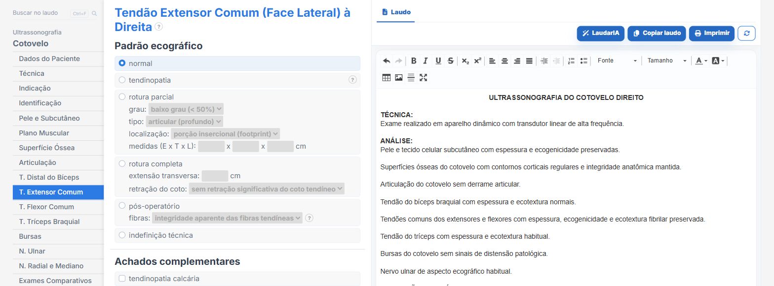
In the elbow template of Laudário, with laterality:
- Tendons, ligaments, ulnar nerve and bursae organized by compartment.
- The conclusion is built from the findings checked.
- Recommendations in their own tab.
Try it for 15 days free, no credit card.
The ultrasound reporting platform built by sonologists. We write about reports, classifications and standardization from the real routine of the practice. Who writes and how.
Educational content for healthcare professionals. It does not replace clinical judgment or the guidelines adopted by your service. Editorial policy.