Peripheral nerve ultrasound: carpal tunnel syndrome and the value of cross-sectional area
Cross-sectional area is the core of peripheral nerve ultrasound, but the absolute value rarely suffices. See the cutoffs by nerve and site, the wrist-to-forearm ratio, the honeycomb pattern, reading the innervated muscle and the causes that only ultrasound finds.
 LaudárioUltrasound reports in practice
This article is part of the guide Musculoskeletal Ultrasound: The Complete Guide, Joint by Joint.
Carpal tunnel syndrome is the most common compressive neuropathy in the human body, and for decades its diagnosis depended on an exam that patients detest: electromyography. Ultrasound has changed this scenario quietly and decisively. Today, the main guidelines recognize ultrasonography as a diagnostic tool of value comparable to neurophysiologic testing for confirming CTS, with three advantages that electrodiagnostic testing lacks: it is painless, it shows the cause of the compression and it detects the anatomical variants that change surgery.
The core of this exam is a single measurement: the nerve's cross-sectional area (CSA). But measuring it well, and above all interpreting it well, requires understanding why an absolute number rarely suffices, and why segmental comparison within the same nerve usually says more than any reference table.
What ultrasound sees that electromyography does not
Electrodiagnostic testing measures function: how long the impulse takes to cross the compressed segment. It is excellent data, and it remains the reference. But it is blind to anatomy. It tells you that the median nerve is compressed at the wrist; it does not tell you what is compressing it.
Ultrasound answers exactly the complementary question. It shows the ganglion cyst pushing on the nerve, the bifid median nerve variant, the persistent median artery, the lumbrical muscle with an anomalous origin, the flexor tenosynovitis occupying the tunnel, amyloid material, the nerve sheath mass. It shows recurrent subluxation of the ulnar nerve over the medial epicondyle during elbow flexion, a diagnosis that exists only on dynamic examination and that electrodiagnostic testing will never make. And in patients with normal electrodiagnostic testing but a convincing clinical picture, it often finds what the functional study did not capture.
There is also a scenario in which ultrasound is irreplaceable: nerve trauma. In the first weeks after an injury, electrodiagnostic testing cannot distinguish neurapraxia from complete transection, since both show no response. Ultrasound shows, immediately, whether the nerve is anatomically continuous or transected. It is the difference between observing and operating urgently.
Cross-sectional area: how to measure
CSA is measured in the transverse view, with the transducer perfectly perpendicular to the course of the nerve, tracing the nerve just inside the inner margin of the hyperechoic epineurium, and not outside it. It is a small difference in the trace that produces relevant differences in the result, and it is the most common source of disagreement between examiners.
Two other sources of error deserve attention:
- Anisotropy. A minimal tilt of the transducer makes the nerve look hypoechoic and artificially widens its outline. The plane must be strictly orthogonal.
- Transducer pressure. Compressing flattens the nerve and underestimates the area. Measure with abundant gel and minimal pressure.
The exam is performed with a high-frequency linear transducer (typically 7 to 18 MHz), in transverse and longitudinal planes, and the scan is dynamic: the nerve is followed along its entire accessible length, not only at the suspected site.
Reference values by nerve and site
CSA varies by nerve and by level, which is why a table is indispensable. The values below are among the most used in practice:
Median nerve
- Wrist (carpal tunnel inlet): up to about 10 mm²
- Forearm (pronator quadratus, 12 cm proximal to the tunnel): up to about 9 mm²
- Mid forearm: up to about 9 mm²
- Antecubital fossa: up to about 11 mm²
- Mid arm: up to about 11 mm²
Ulnar nerve
- Guyon canal (wrist): up to about 6.5 mm²
- Pre-cubital tunnel (forearm): up to about 7.5 mm²
- Cubital tunnel (elbow): up to about 8 mm²
- Mid arm: up to about 9.5 mm²
Other frequent sites
- Radial nerve at the spiral groove of the humerus: up to about 8 mm²
- Common fibular (peroneal) nerve in the lateral popliteal fossa: up to about 17 mm²; at the fibular head: up to about 15 mm²
- Tibial nerve in the popliteal fossa: up to about 32.5 mm²; at the ankle (medial malleolus): up to about 21.5 mm²
- Sural nerve in the leg (10 cm proximal to the lateral malleolus): up to about 6.3 mm²
- Cervical roots: C5 up to about 8 mm²; C6 up to about 8.5 mm²; C7 up to about 8 mm²
An honest caveat about these numbers: the cutoffs in the literature vary. For the median nerve at the wrist, values between 8.5 and 13 mm² are found depending on the study, the population and the tracing method. That is why the absolute value, on its own, is the most fragile parameter of the exam, and why comparisons exist.
What really decides: segmental comparison
Here is the central concept of this article. A compressed nerve does not swell uniformly: it swells focally, just before and at the point of constriction, while the proximal segment remains normal. It is this gradient, and not the absolute value, that characterizes compressive neuropathy.
The practical consequence is large: intranerve comparison normalizes the patient against himself. A large 110 kg man has bigger nerves than a 50 kg woman, and neither reads well against a population table. But each person's wrist/forearm gradient is comparable.
Carpal tunnel: the two ways to measure the gradient
There are two established approaches, and it is worth knowing both:
- Wrist-to-forearm ratio (WFR): the wrist CSA is divided by the forearm CSA (pronator quadratus, 12 cm proximal). A ratio ≥ 1.4 is the classically described cutoff, with high sensitivity and specificity, often superior to those of the absolute value alone.
- Segmental difference (ΔCSA): the forearm CSA is subtracted from the wrist CSA. A difference greater than 2 mm² characterizes focal enlargement.
Both measure the same phenomenon using different arithmetic. A practical decision ladder, combining absolute value and gradient, works like this:
- Wrist CSA above 14 mm²: compressive neuropathy. The absolute value is already sufficient; the gradient is confirmatory.
- Wrist CSA between 8 and 14 mm² with Δ greater than 2 mm²: focal compression in the tunnel. This is where segmental comparison does all the work: the absolute value alone would be ambiguous.
- Wrist CSA between 8 and 14 mm² without a significant difference: borderline. The report should say so, rather than force a conclusion.
- Wrist CSA below 8 mm²: normal.
Note what the intermediate range means: most real patients fall into it. Measuring only the wrist, in this range, is flipping a coin. Measuring the forearm takes thirty seconds and solves it.
Gradients at other sites
- Ulnar nerve at the cubital tunnel: the CSA at the tunnel is compared with the pre-tunnel CSA; a difference greater than 2 mm² characterizes focal enlargement.
- Common fibular nerve at the fibular head: here the comparison is percentage-based: an increase of 20% or more relative to the proximal segment (lateral popliteal fossa) indicates focal enlargement.
Bilateral asymmetry
There is a third axis of comparison, especially useful when the disease is asymmetric: the bilateral ΔCSA, measuring the same site on both sides.
- Δ up to 2 mm²: no significant asymmetry.
- Δ above 2 and up to 4 mm²: moderate asymmetry.
- Δ above 4 mm²: marked asymmetry.
This axis supports leprosy screening, a disease in which asymmetric nerve thickening is a diagnostic criterion and in which ultrasound plays an important and underused role in Brazil. Keep in mind that CTS is also frequently bilateral, which reduces the usefulness of contralateral comparison in this specific context: if both wrists are compressed, symmetry is not reassuring.
Beyond CSA: the internal architecture of the nerve
Reducing the exam to area wastes the method. The normal nerve has a very characteristic appearance in the transverse view, the honeycomb pattern: individual hypoechoic fascicles separated by hyperechoic epineurium and perineurium. It is one of the most recognizable patterns in musculoskeletal ultrasonography, and its deterioration can be graded.
Echogenicity
The comparison standard is the adjacent muscle. The compressed nerve progressively loses echogenicity from endoneurial edema: slightly reduced, moderately reduced, or frankly hypoechoic. Increased echogenicity, on the other hand, suggests fibrosis, as in a chronically injured or postoperative nerve.
Fascicular pattern
The scale is ordinal and worth using precisely:
- Preserved: clear honeycomb.
- Partially preserved.
- Reduced: poorly individualized fascicles.
- Effaced: loss of fascicular definition. This is the finding of greatest structural severity.
- Heterogeneous: asymmetry among the fascicles, which may indicate selective involvement.
Signs of compression in the longitudinal plane
The longitudinal view shows what the transverse view does not: the notch sign, an abrupt constriction of the nerve at the exact point of entrapment, with fusiform dilation immediately proximal. It is the finding that localizes the compression with millimetric precision. In the carpal tunnel, it appears under the flexor retinaculum; in the cubital tunnel, under the Osborne ligament.
The picture is completed by focal flattening of the nerve at the compression site, loss of the perineural fat plane and perineural edema.
Doppler: intraneural vascularity
A concept worth fixing in mind: the normal nerve has no detectable intraneural flow. The presence of vascularity within the nerve is, by itself, a pathologic sign. It reflects inflammatory or compressive hyperemia and correlates with symptom severity in CTS. Perineural hyperemia, without intraneural flow, is an intermediate and more nonspecific finding.
It is worth distinguishing three states: physiologic perineural vascularity without intraneural flow (normal); isolated perineural hyperemia; and abnormal intraneural vascularity, the latter always deserving explicit mention in the conclusion.
The innervated muscle: where time shows
This is the section most often missing from reports, and it answers a question that no other answers: how long has this nerve been suffering?
The golden rule, which must be clear to those who report and to those who read: sonographic signs of denervation appear with a latency of weeks after the acute nerve injury. The absence of muscle changes does not exclude acute neuropathy; it only indicates that there has not yet been time.
Reading the muscle has two axes that point to opposite phases:
- Reduced echotexture: muscle edema. It is the early sign of acute denervation, and it is counterintuitive: the muscle darkens before it brightens.
- Increased echotexture: progresses from partial loss of fiber differentiation to fibro-fatty replacement of the parenchyma. It is the sign of chronic denervation.
- Reduced volume: atrophy, gradable from mild to marked, also a marker of chronicity.
In the carpal tunnel, the target is the thenar eminence (abductor pollicis brevis). An atrophic, hyperechoic thenar in a patient with CTS changes the conversation: it is no longer a case for a night splint, it is a case for decompression, and motor recovery may already be compromised. This information is worth more for the clinical decision than the CSA itself.
Focal findings: when the compression has a culprit
Not every compressive neuropathy is idiopathic, and ultrasound is the only routine method that finds the cause. It is worth actively looking for:
- Ganglion cyst: anechoic, with posterior acoustic enhancement, and the decisive finding: connection to a tendon or joint structure. It is a classic cause of ulnar neuropathy at the Guyon canal and of fibular neuropathy at the fibular neck.
- Nerve sheath tumor (schwannoma or neurofibroma): a solid lesion in continuity with the course of the nerve. The schwannoma tends to be eccentric, displacing the fascicles; the neurofibroma, central. The distinction matters surgically, because a schwannoma can be enucleated while preserving the nerve.
- Malignant peripheral nerve sheath tumor (MPNST): rapid growth, irregular or infiltrative margins, marked hypervascularity. It warrants contrast-enhanced MRI and referral.
- Neuroma: in continuity (the nerve maintains continuity, with fusiform thickening at the injury site) or terminal (at the end of a transected nerve). The distinction defines the repair technique.
- Anatomical variants: bifid median nerve, persistent median artery, fibrous bands. These findings change surgery and are not seen by electrodiagnostic testing. An undescribed bifid median nerve is a surgical risk transferred to the patient.
- Extrinsic compressive structures, lipomas, perineural hematomas and flexor tenosynovitis occupying the tunnel.
Two complementary signs are worth recording whenever present: the mass effect on the nerve, with displacement and compression of the bundle, and the sonographic Tinel sign: pressure with the transducer over the lesion reproduces the symptoms in the nerve's territory. The latter is the direct bridge between the image and the complaint, and it is free.
Pitfalls in the peripheral nerve report
- Measuring only the wrist: in the intermediate range, where most patients fall, the absolute value alone decides nothing.
- Tracing outside the epineurium: systematically overestimates the CSA.
- Compressing with the transducer: flattens the nerve and underestimates the area.
- Ignoring anisotropy: an oblique plane creates a hypoechoic, widened nerve that does not exist.
- Concluding there is no neuropathy because the muscle is normal: denervation takes weeks to appear.
- Not assessing the thenar in CTS: atrophy is what changes surgical urgency.
- Not describing anatomical variants: bifid median nerve and persistent median artery are surgical information.
- Forgetting the dynamic ulnar exam: subluxation over the medial epicondyle exists only in flexion.
- Comparing sides in bilateral CTS: symmetry, here, is not reassuring.
- Reporting “thickened nerve” without the measurement and without the gradient: that is an impression, not an exam.
How Laudário helps structure the peripheral nerve report
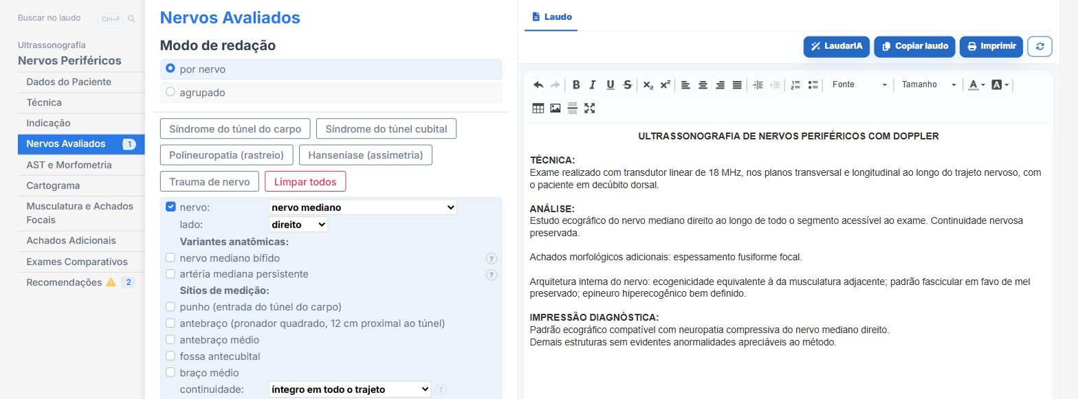
Laudário has a dedicated peripheral nerve module that treats CSA for what it is: a measurement that only means something in comparison. In practice, this means:
- Thirty-seven selectable nerves, from the upper limb, brachial plexus, lower limb and other territories, with predefined measurement sites and the reference value displayed next to each field.
- Automatic calculation of the intranerve gradient: wrist-forearm difference for the median nerve, tunnel-pre-tunnel for the ulnar nerve and percentage increase at the fibular head, each compared with its threshold, with no manual arithmetic in the middle of the exam.
- Automatic calculation of the bilateral ΔCSA with asymmetry classification, which is the axis of leprosy screening and asymmetric neuropathies.
- A morphometry summary table derived automatically from the panels, with the status of each site marked against its reference.
- One-click templates for routine scenarios (carpal tunnel syndrome with bilateral median nerves, wrist and proximal forearm; cubital tunnel; polyneuropathy screening; leprosy; and nerve trauma) that already set up the correct nerves and sites.
- Dedicated fields for internal architecture: echogenicity, fascicular pattern, epineurium, calcifications and Doppler, with phrases that describe the honeycomb and its deterioration on an ordinal scale.
- An innervated muscle tab with acute versus chronic logic built in (reduced echotexture concludes acute denervation; markedly increased echotexture or marked atrophy conclude chronic denervation), and with the latency caveat recorded in the module itself.
- A dynamic adder for focal findings with nature, location, measurements, Doppler and complementary signs (joint connection, mass effect, sonographic Tinel), covering ganglion cyst, neuromas, sheath tumors and variants.
- Concise, descriptive conclusions, without raw numeric jargon, and recommendations auto-suggested according to the finding: clinical correlation and complementary electrodiagnostic testing for abnormalities, specialist referral for discontinuities and masses, contrast-enhanced MRI for expansile lesions.
- Smart omission: the gradient is not printed when the exam is normal, because a normal report does not need arithmetic; it needs to be short and clear.
- Support from LaudarIA, the built-in artificial intelligence, to help write complex cases, such as polyneuropathies with multiple sites or postoperative follow-ups.
The result is a report that answers the neurologist's question and the hand surgeon's question at the same time: is there compression, exactly where, to what degree, from what cause, and for how long. In a condition as prevalent as carpal tunnel syndrome, and in an exam that is still performed by few services with a rigorous method, offering this level of structure is a real differentiator in your region.
If you want to start doing peripheral nerve ultrasound, or already do it and build the report manually, it is worth trying: Laudário offers a free 15-day trial, no credit card required, with immediate access to the peripheral nerve module, the musculoskeletal reports for wrist, hand, elbow, shoulder and knee, and all the other routine protocols.
The ultrasound reporting platform built by sonologists. We write about reports, classifications and standardization from the real routine of the practice. Who writes and how.
Educational content for healthcare professionals. It does not replace clinical judgment or the guidelines adopted by your service. Editorial policy.