Baker cyst on ultrasound: the sign that confirms it, rupture and what is not a cyst
The neck between the semimembranosus and the medial gastrocnemius, the ruptured cyst that mimics thrombosis, the popliteal aneurysm that must not be mistaken for it, and the cause inside the knee.
 LaudárioUltrasound reports in practice
This article is part of the guide Musculoskeletal Ultrasound: The Complete Guide, Joint by Joint.
Baker cyst is one of the most frequent findings on knee ultrasound and one of the most common reasons for an exam request for a "lump behind the knee". Most of the time, it is benign and secondary to some abnormality inside the joint. But there are two moments when the report makes a real difference: when it ruptures and mimics a venous thrombosis, and when a mass behind the knee is not, in fact, a cyst.
This text shows how to confirm the diagnosis, what to describe and how to differentiate the ruptured cyst from thrombosis.
What it is
A Baker cyst (or popliteal cyst) is distension of the bursa between the medial gastrocnemius and the semimembranosus, which communicates with the knee joint. Any condition that increases joint fluid (osteoarthritis, meniscal tear, rheumatoid arthritis, gout) can fill it. For this reason, the cyst is almost always a marker of intra-articular disease, and not the disease itself.
The sign that confirms it: the neck between the tendons
The sonographic diagnosis is not just a "collection in the popliteal fossa". The characteristic finding is the neck of the cyst passing between the semimembranosus tendon and the medial head of the gastrocnemius, on an axial view of the posteromedial aspect of the knee. Seeing this neck confirms that the collection is a Baker cyst and not another lesion.
The report describes:
- Measurements in all three axes, to allow comparison.
- Content: anechoic, with debris, with septations, with loose bodies or synovitis (synovial thickening with flow on Doppler).
- Extension: large cysts can dissect into the calf.
- Associated joint effusion, in the suprapatellar recess.
Ruptured cyst: the differential with thrombosis
When the cyst ruptures, synovial fluid leaks between the calf muscles and causes pain, swelling and warmth, a clinical picture very similar to that of deep vein thrombosis, the so-called pseudothrombophlebitis. Ultrasound resolves both at once:
- Ruptured cyst: loss of the rounded contour, with a pointed inferior border, and fluid dissecting between the medial gastrocnemius and the soleus, in a sheet-like pattern.
- Deep veins: assess the compressibility of the popliteal and calf veins. A ruptured cyst and thrombosis can coexist, and a large cyst can compress the popliteal vein.
For this reason, in a patient with calf pain and a Baker cyst, the report should make clear whether the veins were assessed and are patent. A ruptured cyst described without a venous assessment leaves the most important question unanswered. See our text on venous Doppler for suspected DVT.
When it is not a Baker cyst
Not every mass in the popliteal fossa is a cyst. The main differentials:
- Popliteal artery aneurysm: pulsatile structure, with flow on Doppler and mural thrombus. Often bilateral and associated with an aortic aneurysm. Mistaking it for a cyst is the most serious error, and color Doppler prevents it.
- Meniscal (parameniscal) cyst: next to the meniscus, usually with an associated meniscal tear, lateral or medial.
- Ganglion cyst: no communication with the typical bursa.
- Soft tissue tumors, such as synovial sarcoma, which can have cystic areas and look benign. A solid component with flow on Doppler, without the characteristic neck, calls for investigation with MRI.
The cause inside the knee
Since the cyst is secondary, the report gains value when it looks for the cause within reach of ultrasound: joint effusion, synovitis, osteophytes and signs of osteoarthritis, extrusion of the medial meniscus and crystal deposits in the cartilage. Ultrasound does not assess the interior of the meniscus and the cruciate ligaments well; when clinical suspicion is of an internal derangement, it is worth recommending MRI.
Common mistakes
- Calling any posterior collection a Baker cyst without seeing the neck.
- Not using Doppler and missing a popliteal aneurysm.
- A ruptured cyst without assessment of the deep veins.
- Omitting the measurements, which prevents follow-up.
- Not mentioning the associated effusion and synovitis.
See also our text on the musculoskeletal knee report.
How Laudário helps
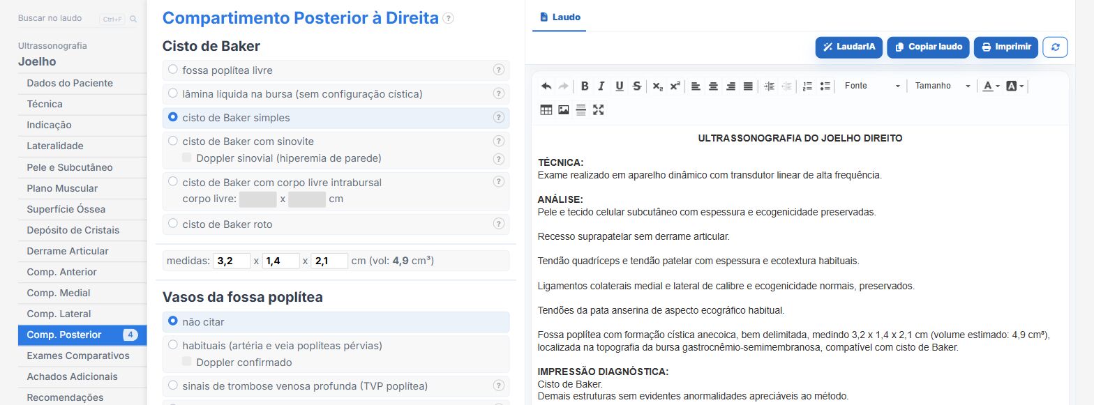
In the Laudário knee template, with laterality:
- Baker cyst with measurements, content and signs of rupture.
- Joint effusion and synovitis in their own fields.
- Venous Doppler templates to complete the exam when thrombosis is suspected.
- Recommendations in their own tab.
Try it for 15 days free, no credit card required.
The ultrasound reporting platform built by sonologists. We write about reports, classifications and standardization from the real routine of the practice. Who writes and how.
Educational content for healthcare professionals. It does not replace clinical judgment or the guidelines adopted by your service. Editorial policy.