Knee ultrasound: how to structure the musculoskeletal report from tendons to Baker cyst
How to structure the knee ultrasound report clearly: tendons, collateral ligaments, joint recesses, Baker cyst, and dynamic maneuvers.
 LaudárioUltrasound reports in practice
This article is part of the guide Musculoskeletal Ultrasound: The Complete Guide, Joint by Joint.
Knee ultrasound has earned a permanent place in the musculoskeletal workup. It is a fast, dynamic exam with no radiation that, in trained hands, can assess structures once reserved for MRI: the patellar and quadriceps tendons, collateral ligaments, synovial recesses, the prepatellar bursa, the popliteal cyst, and even dynamic maneuvers for patellofemoral impingement and subluxation. Precisely because of this versatility, the report needs to be organized by compartment and follow a reproducible sequence. Otherwise, important findings get diluted in the text and the referring physician wastes time looking for the information that matters.
Why standardize the knee ultrasound report?
The knee is a joint with many structures in a small space, examined in four compartments (anterior, medial, lateral, and posterior) with its own dynamic maneuvers. Without a defined structure, the report becomes a loose list of findings in which patellar tendinopathy, joint effusion, and Baker cyst appear mixed together, with no clinical hierarchy.
The main problems that a lack of standardization causes in this exam are:
- Findings in the wrong compartment: describing the Baker cyst in the middle of the patellar tendon paragraph, or citing the medial collateral ligament together with the quadriceps tendon, makes comparative reading harder.
- Inconsistent terminology: alternating between “tendinosis,” “tendinopathy,” and “tendinitis” within the same practice, or describing the same effusion sometimes as “small amount of fluid” and sometimes as “small-volume joint effusion,” confuses follow-up.
- No objective measurements: not reporting the patellar tendon thickness, the estimated volume of the Baker cyst, or the depth of the suprapatellar recess takes away the treating physician's reference for future follow-up.
- Forgotten dynamic maneuvers: failing to mention dynamic assessment of the suprapatellar recess, of the medial collateral ligament course under valgus stress, or of patellofemoral impingement reduces the value of the exam.
- Generic diagnostic impression: concluding only “degenerative changes” or “nonspecific exam,” without returning to the findings that justify the conclusion.
Recommended structure for the knee ultrasound report
The safest approach is to organize the report by the four classic compartments of the exam, always keeping the same order between patients. This makes typing, comparative reading, and the clinic's internal audit easier. We suggest the following blocks:
1. Identification and technique
Include the type of exam (right knee, left knee, or bilateral comparison), the positioning used (supine and prone), the transducer used, and any technical limitations: large effusion that limits the acoustic window, obesity, or pain that limits flexion. This block gives context to the requesting physician and protects the examiner in technically limited exams.
2. Anterior compartment
Describe, in short paragraphs and in the same order, the quadriceps tendon (thickness, echotexture, fibrillar pattern, presence of calcifications or enthesopathy at the patellar insertion), the patellar tendon (origin at the inferior pole, body, and tibial insertion, thickness in the proximal third), the prepatellar and deep infrapatellar bursae, Hoffa's fat pad, and the suprapatellar recess (presence, estimated volume, and fluid characteristics, synovial thickening, loose bodies). In patients with anterior complaints, it is worth recording dynamic assessment with quadriceps contraction.
3. Medial compartment
Assess the medial collateral ligament along its entire length (superficial and deep portions), the medial joint line (osteophytes, peripheral meniscal bulging, parameniscal cyst), the pes anserinus, and the anserine bursa. When indicated, describe the dynamic maneuver under valgus stress. A description of meniscal extrusion beyond the joint margin should always be interpreted with caution and correlated with the clinical picture and other methods.
4. Lateral compartment
Include the lateral collateral ligament, the iliotibial band and its insertion at Gerdy's tubercle, the biceps femoris tendon, the posterolateral corner complex, and the lateral joint line. Lateral parameniscal cysts and iliotibial bursitis are common findings that should have their own paragraph, with location and measurements.
5. Posterior compartment
Describe the popliteal fossa with special attention to the Baker cyst (presence, dimensions in three planes, estimated volume, anechoic content or content with debris, communication with the joint, signs of rupture). Also assess the hamstring tendons, the popliteal neurovascular bundle (ruling out associated deep vein thrombosis when the complaint is calf swelling), and the medial head of the gastrocnemius.
6. Diagnostic impression and suggested management
The conclusion should be objective, hierarchical, and directly correlated with the descriptive body. Start with the findings of greatest clinical relevance (tendon tear, large joint effusion, ruptured Baker cyst), continue with degenerative findings, and finish with incidental findings. Whenever pertinent, suggest clinical correlation or a complementary MRI study for detailed intra-articular assessment (menisci, cruciate ligaments, cartilage). The final decision belongs to the treating physician, but the report should provide every element needed to make it.
Common errors in the knee ultrasound report
Even with a defined structure, some recurring errors undermine the quality of knee reports:
- Confusing the quadriceps and patellar tendons in measurements: swapping the thickness reported in the conclusion is more common than it seems and directly affects the diagnosis of tendinopathy.
- Not measuring the Baker cyst: describing it only as “present” loses the chance of comparison in future exams and of guiding management (aspiration, injection, conservative).
- Underestimating the joint effusion: citing a “small fluid layer” in the suprapatellar recess without reporting its objective depth makes follow-up difficult.
- Forgetting dynamic maneuvers: not assessing the course and stability of the tendons in flexion and extension significantly reduces the diagnostic value of the method.
- Diagnosing a meniscal tear by ultrasound: establishing an intra-articular meniscal tear by ultrasound is a classic trap. The method assesses the peripheral third and parameniscal cysts well, but MRI remains the standard for intrasubstance tears.
- Conclusion disconnected from the description: concluding “exam without abnormalities” when the body of the report describes patellar tendon thickening and a small Baker cyst, or vice versa.
How Laudário helps structure knee reports
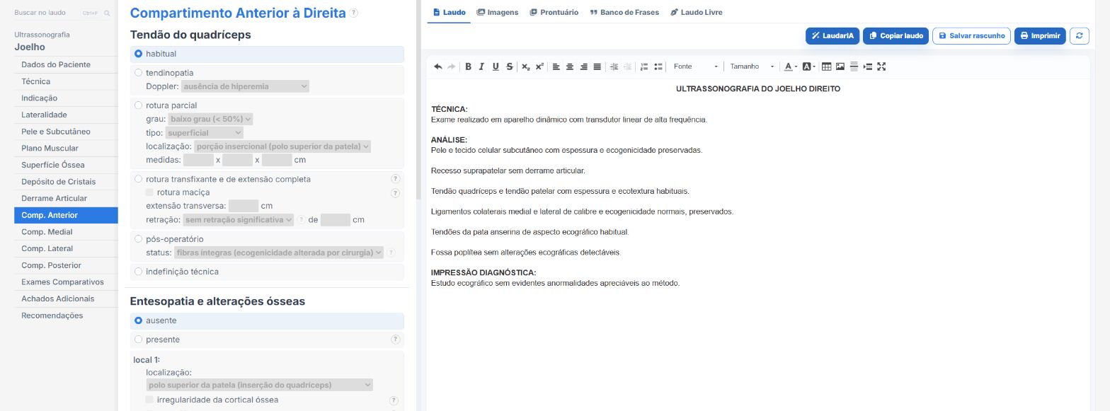
Laudário was designed for professionals who face this routine every day. For knee ultrasound, and for the main musculoskeletal protocols, the system offers:
- Ready-made, structured templates for the knee organized by the four compartments (anterior, medial, lateral, and posterior), in the classic sequence of the exam, to speed up typing without missing any structure.
- Standardized, customizable phrases for describing tendinopathies, joint effusion, Baker cyst, enthesopathies, and bursitis, keeping terminology consistent between reports and between professionals.
- Integrated calculations and measurements in the typing workflow (tendon thickness, popliteal cyst dimensions, recess depth), avoiding manual math and reducing transcription errors.
- A complete library covering musculoskeletal exams (shoulder, knee, ankle, elbow, wrist), as well as abdomen, pelvis, small parts, obstetrics, and Doppler, all in a single online system, which makes standardization between professionals in the same clinic easier.
The result is simple: less time typing, fewer forgotten findings, and more consistent reports across exams of the same patient, which makes all the difference in musculoskeletal assessment, where follow-up of tendinopathies and effusions is central to management. Instead of starting from a blank page with every patient, you adjust a template that has already been tested, keep the clinic's professional standard, and focus on what really demands your attention: reading the images.
If you still build your knee reports manually or by copying old text, it is worth getting to know Laudário. Try it free for 15 days and see in practice how a well-designed structure saves time on every exam.
The ultrasound reporting platform built by sonologists. We write about reports, classifications and standardization from the real routine of the practice. Who writes and how.
Educational content for healthcare professionals. It does not replace clinical judgment or the guidelines adopted by your service. Editorial policy.