Hand and Wrist Ultrasound: De Quervain, Trigger Finger, Ganglion Cyst and Carpal Tunnel
The four complaints behind most hand and wrist ultrasound requests and what the report needs for each: the De Quervain septum, the A1 pulley, the origin of the cyst and the median nerve area.
 LaudárioUltrasound reports in practice
This article is part of the guide Musculoskeletal Ultrasound: The Complete Guide, Joint by Joint.
The hand and wrist pack dozens of tendons, pulleys, ligaments, nerves and joints into a few centimeters. It is precisely this density that makes ultrasound the method of choice for most complaints: high spatial resolution, dynamic assessment with the patient moving the painful finger, and immediate comparison with the opposite side.
The problem usually lies in the report. "Flexor tenosynovitis" without saying which finger, which pulley and what thickness; "ganglion cyst" without the origin; "normal carpal tunnel" without the nerve area. This text organizes the four complaints that account for most requests (De Quervain, trigger finger, ganglion cyst and carpal tunnel) and what each one needs written to be useful to the hand surgeon and the rheumatologist.
How to organize the exam
The safest way not to forget structures is to follow the anatomy by compartments, always in the same order:
- Dorsal wrist: the six extensor compartments, from radial (I) to ulnar (VI), with Lister's tubercle as the landmark between II and III.
- Volar wrist: carpal tunnel (median nerve and nine flexor tendons), Guyon's canal (ulnar nerve and artery) and the flexor carpi radialis tendon.
- Fingers: flexor digitorum superficialis and profundus tendons, annular pulleys (A1 to A5) and the extensor apparatus.
- Joints: radiocarpal, intercarpal, metacarpophalangeal and interphalangeal, with attention to effusion and synovitis.
- Ligaments: dorsal scapholunate and, in the thumb, the ulnar collateral ligament of the metacarpophalangeal joint.
A report that follows this order is easier to read and, above all, makes clear what was evaluated. "Remaining extensor compartments unremarkable" tells the referring physician that they were looked at.
De Quervain tenosynovitis
This is tenosynovitis of the first extensor compartment, where the abductor pollicis longus and extensor pollicis brevis run over the radial styloid process. Clinically it is radial-sided wrist pain that worsens with the Finkelstein test, common in postpartum women and in people who use the thumb heavily.
The sonographic findings:
- Retinacular thickening of the first compartment, hypoechoic, often with hyperemia on Doppler.
- Tendon thickening and fluid in the sheath.
- Comparison with the contralateral side, which helps a lot in subtle cases.
The detail that adds the most value to the report is the intracompartmental septum. In a relevant proportion of people, a fibrous septum divides the first compartment into two subcompartments. The presence of the septum changes the guided injection (both sides must be reached) and the surgery (both tunnels must be released). Writing "intracompartmental septum present" or "no septum identified" is information the surgeon uses.
Trigger finger
Trigger finger is a conflict between the flexor tendon and the A1 pulley, at the level of the metacarpal head. Ultrasound shows:
- Thickened, hypoechoic A1 pulley, often with hyperemia. Measuring the pulley thickness, compared with another finger or the opposite side, makes the finding objective.
- Flexor tendon thickening just proximal to the pulley, sometimes with a tendon nodule.
- Fluid in the synovial sheath.
- Dynamic assessment: asking the patient to flex and extend the finger shows the tendon catching as it passes through the pulley. When present, it should be in the report.
Always identify the finger (thumb, index, middle, ring, little) and the hand. It seems obvious, but "stenosing flexor tenosynovitis" without the finger forces the referring physician to go back to the exam.
Ganglion cyst
This is the most common mass of the hand and wrist. On ultrasound, it appears as an anechoic or hypoechoic, well-defined, sometimes multiloculated formation, with no flow on Doppler and with posterior acoustic enhancement.
What turns "ganglion cyst" into a useful report is the origin:
- Dorsal: most arise from the scapholunate ligament. Looking for and describing the pedicle (the "neck" connecting the cyst to the joint) is what guides resection; removing the cyst without the pedicle favors recurrence.
- Volar: often next to the radial artery, between it and the flexor carpi radialis. The relationship to the artery must be written before any aspiration.
- Tendon sheath cyst: small, firm, at the base of the fingers, next to the pulleys.
- Occult cyst: small, dorsal, not palpable, and explains pain without a mass. Ultrasound is the method that finds it.
Describe the measurements in three axes, the lobulation and the origin. And remember the differential: giant cell tumor of the tendon sheath is solid and usually has flow on Doppler; a cyst does not.
Carpal tunnel syndrome
This is the most common compressive neuropathy of the upper limb. Ultrasound does not replace electroneuromyography, but it adds what the latter cannot see: the anatomic cause and the morphology of the nerve.
The central parameter is the cross-sectional area of the median nerve at the tunnel inlet, at the level of the pisiform bone. Cutoff values vary between publications; areas above approximately 10 to 12 mm² are considered enlarged, and a wrist-to-distal-forearm area ratio above 1.4 reinforces the diagnosis. The report should give the number, not just "thickened nerve".
Besides the area, it is worth describing:
- Loss of the fascicular pattern and hypoechogenicity of the nerve proximal to the retinaculum.
- Bowing of the flexor retinaculum.
- Secondary causes: flexor tenosynovitis, cyst, anomalous muscle, bifid median nerve, persistent median artery. These variants change the surgical approach.
To go deeper into the measurement criteria, read our article on peripheral nerve ultrasound and cross-sectional area.
Synovitis and the rheumatologic view
In patients with arthritis, hand and wrist ultrasound evaluates synovitis (synovial thickening on gray scale and Doppler signal), erosions (cortical defect seen in two planes) and tenosynovitis. The 0 to 3 semiquantitative scores of the OMERACT/EULAR group standardize the description and allow exams to be compared over the course of treatment. For this audience, a report with the grade per joint is worth far more than "signs of synovitis".
Common mistakes in the report
- Not identifying the finger and the hand in trigger finger and tenosynovitis.
- Omitting the septum in De Quervain.
- Cyst without origin, without a pedicle and without the relationship to the radial artery in volar cysts.
- Carpal tunnel without the median nerve area in square millimeters.
- Not comparing with the contralateral side when the finding is subtle.
- Forgetting the dynamic maneuver when the complaint is catching.
How Laudário helps
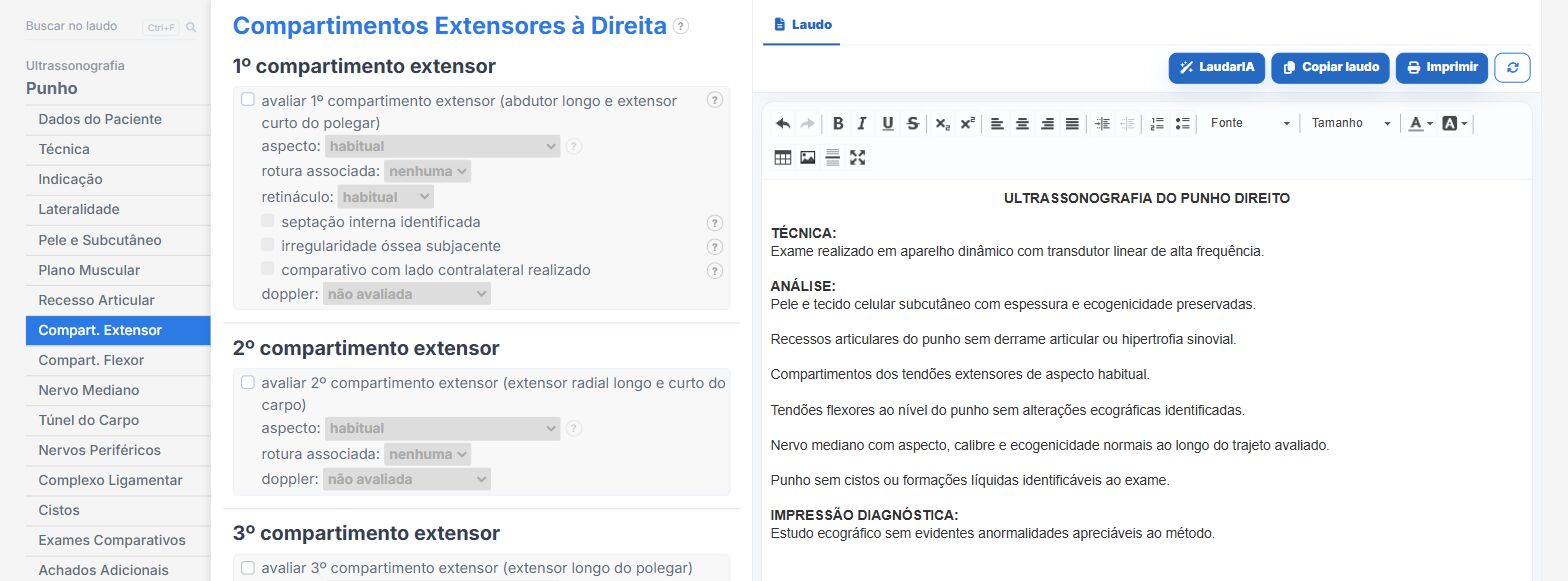
Laudário has dedicated Hand and Wrist modules, as well as the Rheumatologic wrist and hand module and the Peripheral nerves module. In practice:
- Extensor and flexor compartments organized in tabs, with laterality, so that none is left out.
- De Quervain, trigger finger, ganglion cyst and carpal tunnel with ready-made phrases and measurement fields in the right place.
- A median nerve cross-sectional area field, so the number always makes it into the report.
- Recommendations in their own tab, without mixing management with findings.
If you report hand and wrist exams often, it is worth trying: 15 days free, no credit card.
The ultrasound reporting platform built by sonologists. We write about reports, classifications and standardization from the real routine of the practice. Who writes and how.
Educational content for healthcare professionals. It does not replace clinical judgment or the guidelines adopted by your service. Editorial policy.