Ankle and foot ultrasound: Achilles, peroneal tendons, plantar fascia and Morton neuroma
The compartment-based study, insertional and non-insertional Achilles tendinopathy, peroneal longitudinal tears, sprain, plantar fascia thickness and the Morton neuroma space.
 LaudárioUltrasound reports in practice
This article is part of the guide Musculoskeletal Ultrasound: The Complete Guide, Joint by Joint.
The ankle and foot account for very frequent complaints: heel pain on getting out of bed, the thickened Achilles tendon of the runner, the sprain that does not improve, the burning pain between the toes. Ultrasound answers most of them well, with an advantage that MRI does not have: examining the tendon in motion, at the exact site of pain and comparing with the other side.
This text organizes the study by compartments and shows what the report needs to include for each of the most common complaints.
Organization by compartments
Always following the same order avoids missing structures and makes clear, in the report, what was evaluated:
- Anterior: tibialis anterior, extensor hallucis longus and extensor digitorum longus tendons; tibiotalar joint recess (effusion).
- Medial: tibialis posterior, flexor digitorum longus and flexor hallucis longus tendons; tibial nerve in the tarsal tunnel; deltoid ligament.
- Lateral: peroneus brevis and longus tendons; anterior talofibular, calcaneofibular and anterior inferior tibiofibular ligaments.
- Posterior: calcaneal (Achilles) tendon and retrocalcaneal and retro-Achilles bursae.
- Plantar: plantar fascia and intermetatarsal spaces.
Calcaneal (Achilles) tendon
It is the most examined tendon of the foot, and the report needs to state where the abnormality is, because the two forms of tendinopathy have different treatments:
- Non-insertional tendinopathy, about 2 to 6 cm above the insertion: fusiform thickening, loss of the fibrillar pattern, hypoechoic areas and neovascularization on Doppler.
- Insertional tendinopathy, adjacent to the calcaneus: thickening at the insertion, enthesophyte, retrocalcaneal bursitis and, at times, Haglund deformity.
The anteroposterior thickness of the normal tendon is around 5 to 6 mm; values above this, especially compared with the opposite side, support tendinopathy. The measurement should be in the report.
In a tear, the report describes whether it is partial or complete, the distance from the insertion, the size of the gap between the stumps (at rest and in plantar flexion) and the integrity of the plantaris tendon. This information guides the choice between conservative treatment and surgery.
Peroneal tendons
Persistent lateral pain after a sprain often originates in the peroneal tendons. The findings to look for:
- Tenosynovitis: fluid and thickening of the common sheath.
- Longitudinal tear (split) of the peroneus brevis: the tendon appears divided in two, sometimes involving the longus. It is common and underdiagnosed.
- Subluxation or dislocation: seen on the dynamic maneuver of dorsiflexion and eversion, with the tendons leaving the retromalleolar groove. Only the dynamic study shows it.
Posterior tibial tendon
Posterior tibial tendon dysfunction is the main cause of acquired adult flatfoot, more common in women over 50. Ultrasound shows tendinopathy, tenosynovitis or tear, usually in the retromalleolar region and near the insertion on the navicular. It is worth remembering the accessory navicular bone, which can be the cause of medial pain.
Sprain and lateral ligaments
The anterior talofibular ligament is the most injured in an inversion sprain, followed by the calcaneofibular. The report describes:
- Intact, thickened (old or partial injury) or torn ligament.
- Partial or complete injury, and the location (fibular, middle, talar).
- Stability on the anterior drawer maneuver, when performed.
The anterior inferior tibiofibular ligament deserves attention: its injury (high ankle sprain) has a longer recovery.
Plantar fascia
Plantar fasciopathy is the most common cause of heel pain. The main finding is thickening of the fascia at the calcaneal insertion: above about 4 mm is considered increased, and comparison with the opposite side helps. Hypoechogenicity, loss of the fibrillar pattern and perifascial fluid complete the picture. A calcaneal spur may be present, but it is not the cause of the pain by itself.
It is also worth looking for partial tear of the fascia, which can occur spontaneously or after injection, and plantar fibromatosis nodules (Ledderhose disease), in the middle third of the fascia.
Morton neuroma
Burning pain radiating to the toes, with a sensation of a "pebble in the shoe", suggests Morton neuroma: a perineural fibrosis of the digital nerve, most common in the third intermetatarsal space (between the third and fourth metatarsals), followed by the second.
- Hypoechoic, oval image in the intermetatarsal space, oriented along the axis of the nerve.
- Sonographic Mulder sign: with lateral compression of the forefoot, the neuroma is displaced plantarward, sometimes with a click.
- Frequently associated with intermetatarsal bursitis.
The report should state the space involved, the measurements and the presence of bursitis. Small neuromas, below 5 mm, may not explain the symptoms.
Common errors
- Achilles tendinopathy without saying whether it is insertional or non-insertional.
- Tear without measuring the gap between the stumps.
- Lateral pain without dynamic evaluation of the peroneal tendons.
- Plantar fascia without the thickness.
- Neuroma without the intermetatarsal space.
For the same logic applied to the knee, see our text on the musculoskeletal knee report.
How Laudário helps
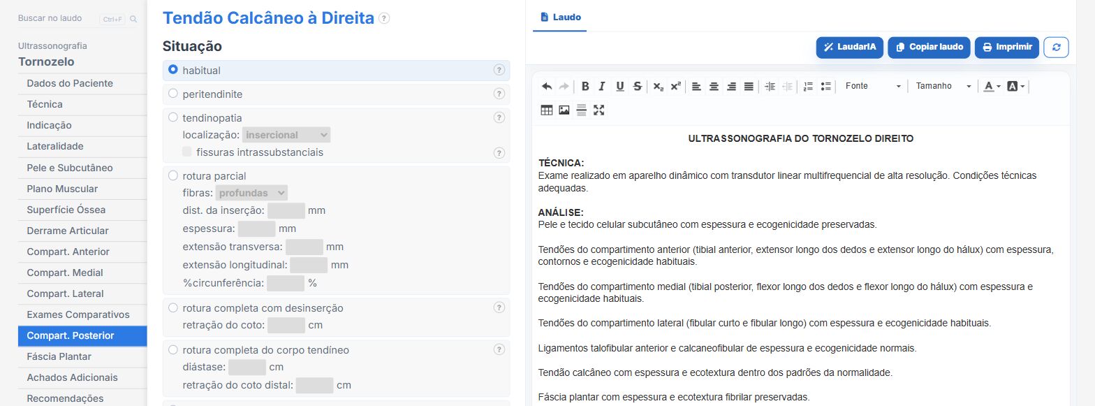
Laudário has dedicated Ankle and Foot templates, with laterality. In practice:
- Compartments organized in tabs, so no tendon is left out.
- Calcaneal tendon with insertional and non-insertional forms and the measurement fields.
- Peroneal tendons, posterior tibial, ligaments, plantar fascia and Morton neuroma with ready-made phrases.
- Recommendations in their own tab.
Try it for 15 days free, no credit card.
The ultrasound reporting platform built by sonologists. We write about reports, classifications and standardization from the real routine of the practice. Who writes and how.
Educational content for healthcare professionals. It does not replace clinical judgment or the guidelines adopted by your service. Editorial policy.