Twin pregnancy: chorionicity, amnionicity, discordance and twin-twin transfusion in the report
Chorionicity is the information that defines all follow-up. The lambda and T signs, consistent fetal identification, weight discordance, Quintero staging and anemia-polycythemia in monochorionic twins.
 LaudárioUltrasound reports in practice
This article is part of the guide Obstetric ultrasound: the three-trimester guide, from dating to Doppler.
In a twin pregnancy, one piece of information is worth more than all the others: chorionicity. It defines the risk, the frequency of studies and which complications to look for. And it can only be determined reliably in the first trimester. A twin report that does not state chorionicity explicitly forces the obstetrician to treat the pregnancy as higher risk, as a precaution, until the end.
This article walks through what the twin report needs to contain from the first to the third trimester, including the specific complications of monochorionic pregnancies.
Chorionicity and amnionicity
Between 11 and 14 weeks, chorionicity is determined by the junction of the intertwin membrane with the placenta:
- Lambda sign (λ): wedge-shaped chorionic tissue penetrating between the membranes. Indicates a dichorionic pregnancy.
- T sign: the thin membrane inserts at a right angle into the placenta, with no chorionic tissue between the layers. Indicates a monochorionic pregnancy.
Two separate placentas also indicate dichorionicity; so do discordant sexes. But an apparently single placenta does not mean monochorionicity, because dichorionic placentas can fuse.
Amnionicity is determined by the presence of the intertwin membrane. When no membrane is seen in a monochorionic pregnancy, it is monoamniotic, the highest-risk form, with cord entanglement.
The report should record the sign observed, the conclusion (dichorionic diamniotic, monochorionic diamniotic or monochorionic monoamniotic) and the gestational age at which the determination was made. If it was not possible to determine, this must be written, because it changes follow-up.
Dating and fetal identification
In twins, the pregnancy is dated by the larger crown-rump length, so as not to underestimate the age in case of early restriction of one of the fetuses.
Fetal identification must be consistent across studies. The most common approach is to describe the position of each fetus (right or left, upper or lower) and the cord insertion into the placenta, naming them fetus 1 and fetus 2 (or A and B) with the same logic in every study. Swapping the identification between one study and another can hide a growth discordance.
Growth discordance
Weight discordance is calculated as the difference between the estimated weight of the larger and the smaller twin, divided by the weight of the larger. Discordance above 20 to 25% is clinically relevant, depending on the protocol adopted, and should be written in the report with the percentage, along with the weight and percentile of each fetus.
In monochorionic twins, selective fetal growth restriction is classified by the umbilical artery Doppler of the smaller fetus (Gratacós types): positive end-diastolic flow (type I), persistent absent or reversed end-diastolic flow (type II) and intermittent absent or reversed end-diastolic flow (type III). The type changes the prognosis and follow-up.
Twin-twin transfusion syndrome
This is the most feared complication of monochorionic diamniotic twins, caused by unbalanced vascular anastomoses in the placenta. One fetus (donor) has little fluid; the other (recipient) has excess. The sonographic diagnosis is based on fluid discordance:
- Recipient with polyhydramnios: deepest vertical pocket above 8 cm (or above 10 cm after 20 weeks, depending on the criterion).
- Donor with oligohydramnios: deepest vertical pocket below 2 cm.
Quintero staging guides severity: stage I (fluid discordance only), II (donor bladder not visible), III (critical Doppler in one of the fetuses), IV (hydrops) and V (demise of one or both). The report should include the deepest pocket of each sac, the bladder of each fetus, the Doppler and the stage.
For this reason, monochorionic pregnancies are followed with ultrasound every two weeks starting at 16 weeks, with measurement of the fluid in each sac at every study.
Twin anemia-polycythemia sequence (TAPS)
Another complication of monochorionic twins, caused by very small anastomoses: one fetus becomes anemic and the other polycythemic, without significant fluid discordance. The diagnosis is made by the middle cerebral artery peak systolic velocity: above 1.5 multiples of the median (MoM) in the anemic fetus and below 1.0 MoM in the polycythemic one, or a difference between the two above 0.5 MoM, according to the most recent criteria. Measurement of the middle cerebral artery PSV in both fetuses should be part of the follow-up of monochorionic twins.
What every twin report needs to contain
- Chorionicity and amnionicity (or "not determined," with the reason).
- Identification and position of each fetus, consistent with previous studies.
- Biometry, estimated weight and percentile of each fetus.
- Weight discordance as a percentage.
- Amniotic fluid of each sac (deepest vertical pocket).
- Placenta(s) and cord insertions.
- Doppler of each fetus, when indicated, including the middle cerebral artery PSV in monochorionic twins.
- Cervix, because of the increased risk of preterm birth.
- Diagnostic impression with chorionicity repeated and the complications identified.
For cervical measurement, see our article on cervical length measurement in obstetric ultrasound.
How Laudário helps
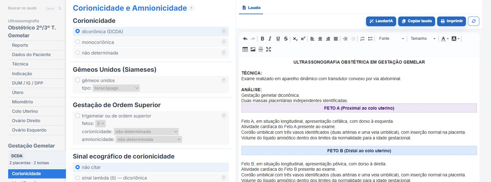
Laudário has twin templates for the first, second and third trimesters, the morphology scan and Doppler, including combinations with the biophysical profile. In practice:
- Chorionicity and amnionicity with the lambda and T signs.
- Biometry and Doppler of each fetus in separate blocks, with weight discordance calculated.
- Twin-twin transfusion and anemia-polycythemia in the monochorionic workflow.
- Diagnostic impression assembled from what was selected, with chorionicity always present.
Try it free for 15 days, with no credit card.
The ultrasound reporting platform built by sonologists. We write about reports, classifications and standardization from the real routine of the practice. Who writes and how.
Educational content for healthcare professionals. It does not replace clinical judgment or the guidelines adopted by your service. Editorial policy.