Placenta on ultrasound: previa, low-lying placenta, accreta and what the grade does not tell you
The distance to the internal os in millimeters, the new terminology for placenta previa and low-lying placenta, signs of accreta in patients with a prior cesarean, the little value of placental grade, and vasa previa.
 LaudárioUltrasound reports in practice
This article is part of the guide Obstetric ultrasound: the three-trimester guide, from dating to Doppler.
The placenta appears in every obstetric report, usually in one line: "anterior placenta, grade I". That line, however, tends to carry the least useful information (the grade) and omit what changes management: the distance to the internal os of the cervix, signs of accreta in patients with a prior cesarean, and the cord insertion.
This text shows what really matters in describing the placenta and how to write each finding.
Location
The placenta is described by the wall of insertion: anterior, posterior, fundal, right or left lateral, or combinations. Location is relevant for cesarean delivery (anterior placenta over the incision region) and for the interpretation of some exams, but the decisive information is something else: the relationship to the cervix.
Placenta previa and low-lying placenta
The old terminology (marginal, partial, total previa) has been replaced by two simpler, more reproducible categories, based on the distance between the placental edge and the internal cervical os, measured by the transvaginal route:
- Placenta previa: the placenta covers the internal os.
- Low-lying placenta: the edge is less than 20 mm from the internal os, without covering it.
The measurement should be in the report in millimeters. The transvaginal route is safe and more accurate than the abdominal one, which overestimates or underestimates the distance depending on bladder filling.
The timing of the diagnosis matters. At the second-trimester anatomy scan, many low-lying placentas "migrate" as the lower segment grows. For this reason, a low-lying placenta or previa at this stage leads to a recommendation for reassessment at around 32 weeks, not to a definitive diagnosis.
Placenta accreta
The placenta accreta spectrum (accreta, increta and percreta) is abnormal invasion of trophoblast into the myometrium, sometimes as far as the serosa and bladder. The main risk factor is the combination of prior cesarean and anterior placenta previa or low-lying placenta, and the frequency has been rising with cesarean rates.
The sonographic signs to look for in these patients:
- Loss of the retroplacental hypoechoic zone (the "clear zone" between the placenta and the myometrium).
- Irregular placental lacunae, with turbulent flow on Doppler ("Swiss cheese" appearance).
- Myometrial thinning below 1 mm.
- Interruption of the uterus-bladder interface, with the placenta bulging toward the bladder.
- Hypervascularity at the uterus-bladder interface and vessels crossing the myometrium.
These signs follow the standardized European terminology and should be described one by one. When accreta is suspected, the report recommends evaluation at a referral center, because delivering a placenta accreta requires a team and planning. One detail: targeted evaluation is only performed when there is a risk factor, and the report should state whether it was done.
Placental grade
The Grannum classification (grades 0 to III) describes maturation by the presence of calcifications. It entered routine reports, but it has little clinical value: grade III placentas are common at term and do not, on their own, indicate placental insufficiency. What assesses placental function is fetal growth, amniotic fluid and Doppler.
The grade can be mentioned, but it should not raise alarm or take the place of relevant information. A grade III placenta before 32 weeks is the exception that deserves attention to growth and Doppler.
Other placental findings
- Placental abruption: ultrasound has low sensitivity. A retroplacental hematoma may be seen, but a normal exam does not exclude abruption, and the report should say so when suspicion is clinical.
- Venous lakes and subchorionic cysts: generally without significance.
- Succenturiate placenta (accessory lobe) and bilobed placenta: important because of the association with vasa previa.
- Increased thickness: associated with hydrops, infections, diabetes and fetal anemia.
Cord insertion and vasa previa
Cord insertion (central, eccentric, marginal or velamentous) should be identified in the second trimester. Velamentous insertion (cord inserted into the membranes) and a placenta with an accessory lobe increase the risk of vasa previa: fetal vessels running in the membranes over the cervix, which rupture with the membranes and cause fetal hemorrhage. With transvaginal color Doppler over the internal os, the diagnosis is simple, and it completely changes management.
Common errors
- Placental grade in place of the distance to the internal os.
- Using marginal, partial and total previa.
- Definitive diagnosis of placenta previa in the second trimester, without recommending reassessment.
- Not looking for accreta in a low anterior placenta with a prior cesarean.
- Cord insertion not evaluated.
See also our text on the second-trimester anatomy scan.
How Laudário helps
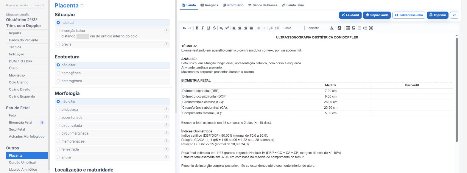
In the obstetric and anatomy scan templates of Laudário:
- Placental location, distance to the internal os and placenta previa or low-lying placenta.
- Signs of accreta as options to check.
- Cord insertion and vasa previa.
- Reassessment recommendation in its own tab.
Try it for 15 days free, no credit card required.
The ultrasound reporting platform built by sonologists. We write about reports, classifications and standardization from the real routine of the practice. Who writes and how.
Educational content for healthcare professionals. It does not replace clinical judgment or the guidelines adopted by your service. Editorial policy.