Obstetric Doppler: umbilical artery, middle cerebral artery, cerebroplacental ratio, and ductus venosus
Each index by percentile for gestational age, the absent end-diastolic flow that cannot be buried in the middle of the text, the cerebroplacental ratio that changes the third trimester, and the ductus venosus that decides delivery.
 LaudárioUltrasound reports in practice
This article is part of the guide Obstetric ultrasound: the three-trimester guide, from dating to Doppler.
Obstetric Doppler is the exam that turns suspected growth restriction into a decision: follow weekly, admit, or deliver early. For that reason, the report cannot be a list of loose indices. For each vessel, it must say whether the value is normal for gestational age, what percentile it falls in, and what that means in the context of the fetus.
This text walks through the four territories that make up the assessment (umbilical artery, middle cerebral artery, cerebroplacental ratio, and ductus venosus), plus the uterine arteries, and shows how to structure the report so the obstetrician finds the information in seconds.
Before the indices: the right gestational age
Every Doppler index is interpreted by percentile for gestational age. If the dating is wrong, the percentile will be wrong. For that reason, the report should make clear which gestational age was used, preferably the one from the first-trimester ultrasound, and not the one estimated from current biometry, which in a growth-restricted fetus will underestimate the age and mask the abnormality.
It is also worth recording the technical conditions: absence of fetal breathing and body movements during the measurement, an insonation angle close to zero for the middle cerebral artery, and the sampling site in the umbilical artery (free loop, fetal insertion, or placental insertion), because values vary along the cord.
Umbilical artery
It reflects placental resistance. The main parameter is the pulsatility index (PI):
- PI above the 95th percentile for gestational age indicates increased placental resistance.
- Absent end-diastolic flow (no diastolic flow) is a serious finding, associated with significant placental insufficiency.
- Reversed end-diastolic flow is the most advanced grade and requires immediate management.
In the report, absent and reversed end-diastolic flow cannot appear as a detail in the middle of a paragraph. They must be described explicitly and repeated in the diagnostic impression.
Middle cerebral artery
It assesses the fetal response to hypoxia. When oxygenation falls, the fetus dilates the cerebral arteries to protect the brain, which lowers the PI of the middle cerebral artery: this is brain sparing, or hemodynamic redistribution.
- PI below the 5th percentile indicates cerebral vasodilation.
- The peak systolic velocity (PSV) has another use: screening for fetal anemia. Values above 1.5 multiples of the median (MoM) for gestational age suggest moderate or severe anemia, for example in alloimmunization or parvovirus infection.
Mixing the two uses is a frequent error. If the indication is anemia, the report must include the PSV in cm/s and in MoM. If it is growth restriction, the focus is the PI.
Cerebroplacental ratio
The cerebroplacental ratio (CPR) is the PI of the middle cerebral artery divided by the PI of the umbilical artery. It combines both sides of the problem: the placenta that resists and the brain that protects itself. A CPR below the 5th percentile is a marker of redistribution and, especially in the third trimester, may be abnormal even when the umbilical and middle cerebral arteries are still individually within limits.
For that reason, the CPR should be calculated and written in every third-trimester Doppler, with its percentile, and not only when the other indices are already abnormal.
Ductus venosus
The ductus venosus shows how the fetal heart is coping with the overload. Its flow has three components, and the most important is the a-wave, which corresponds to atrial contraction:
- Positive a-wave: expected.
- Ductus venosus PI above the 95th percentile: sign of compromise.
- Absent or reversed a-wave: sign of cardiac decompensation, with a high risk of fetal death, which generally leads to early delivery depending on gestational age.
Uterine arteries
They assess the maternal side of the placenta. A mean PI of the two uterine arteries above the 95th percentile indicates inadequate trophoblastic invasion, associated with preeclampsia and growth restriction of placental origin. Persistence of the early diastolic notch after the second trimester complements the description. In the report, record the PI of each side, the mean PI, and the percentile.
Growth restriction: early and late
The international consensus definition of growth restriction (Delphi) separates two forms with different behavior:
- Early, before 32 weeks: usually accompanied by progressive umbilical artery abnormality, followed by the ductus venosus. Umbilical and ductus venosus Doppler guide the timing of delivery.
- Late, from 32 weeks: the umbilical artery is often normal, and the abnormality appears in the middle cerebral artery and the CPR. It is the form most easily missed.
Staging protocols, such as the Barcelona protocol, combine these parameters into stages that guide follow-up frequency and the ideal gestational age for delivery. When the service uses a protocol, citing the stage in the report is very helpful to the obstetrician.
How to structure the report
- Reference gestational age and estimated fetal weight with percentile.
- For each vessel: measured index, percentile, and interpretation (normal, increased, decreased).
- CPR calculated, with percentile.
- Critical findings (absent or reversed end-diastolic flow, absent or reversed a-wave) highlighted and repeated in the impression.
- A diagnostic impression that answers the clinical question: are there or are there not signs of placental insufficiency and hemodynamic redistribution.
- Follow-up recommendation in its own tab, without mixing it with the findings.
For the complete third-trimester assessment, see also our text on well-being, amniotic fluid, and Doppler in the third trimester.
Common errors
- Index without a percentile.
- Percentile calculated with the gestational age from current biometry.
- CPR omitted because the umbilical artery was normal.
- Middle cerebral artery PSV without MoM when the indication is anemia.
- Absent end-diastolic flow described in the middle of the text and missing from the conclusion.
How Laudário helps
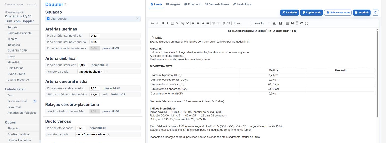
Laudário has obstetric templates with Doppler for the first trimester and for the second and third, including combinations with biophysical profile and twin pregnancies. In practice:
- Index percentiles calculated automatically from the reference gestational age.
- Cerebroplacental ratio calculated and interpreted without hand calculation.
- Absent end-diastolic flow, reversed end-diastolic flow, and ductus venosus abnormalities with their own phrases, which go into the diagnostic impression.
- Recommendations in a separate tab.
Try it for 15 days free, no credit card required.
The ultrasound reporting platform built by sonologists. We write about reports, classifications and standardization from the real routine of the practice. Who writes and how.
Educational content for healthcare professionals. It does not replace clinical judgment or the guidelines adopted by your service. Editorial policy.