Adult hip ultrasound: greater trochanter, gluteal tendons, bursae and joint effusion
Greater trochanteric pain syndrome, gluteus medius and minimus tendinopathy and tears, the bursae, joint effusion and what ultrasound cannot assess in the hip.
 LaudárioUltrasound reports in practice
This article is part of the guide Musculoskeletal Ultrasound: The Complete Guide, Joint by Joint.
Lateral hip pain is one of the most common complaints in orthopedic and rheumatology offices, and the request for a "hip ultrasound for bursitis" is frequent. Most of the time the main problem is not the bursa but the gluteal tendons. A report that describes each structure helps more than the label of bursitis.
Greater trochanteric pain syndrome
This is the name used today for lateral hip pain, because it encompasses several findings that tend to coexist:
- Gluteus medius and gluteus minimus tendinopathy: thickened, hypoechoic tendons with loss of the fibrillar pattern, sometimes with calcifications and irregularity of the trochanteric cortex.
- Tears: partial or complete, with discontinuity of the fibers; in complete tears, retraction of the tendon.
- Bursitis: fluid distension of the subgluteus maximus bursa, the one most often cited as the "trochanteric bursa", or of the gluteus medius and minimus bursae.
Each insertion should be described separately: the facets of the greater trochanter help localize which tendon is involved.
Joint effusion
Effusion is assessed in the anterior recess, with the transducer aligned with the femoral neck. Comparison with the contralateral side is the most useful data: distension of the recess with asymmetry relative to the other side indicates effusion. Ultrasound also guides aspiration when septic arthritis is suspected.
Other findings
- Iliopsoas bursitis: a collection anterior to the hip, which may communicate with the joint.
- Snapping hip: the dynamic exam shows the abrupt sliding of the iliotibial band or gluteus maximus over the trochanter, or of the iliopsoas tendon in the anterior region.
- Hamstring tendinopathy at the ischial tuberosity, a cause of low gluteal pain.
What ultrasound does not assess well
Lesions of the acetabular labrum, cartilage and femoroacetabular impingement are assessed by MRI, with or without arthrography. A normal ultrasound does not exclude these causes, and the report can record this limitation when the clinical indication is mechanical groin pain.
Common mistakes
- Concluding "trochanteric bursitis" without assessing the gluteal tendons.
- Not comparing the anterior recess with the contralateral side.
- Not performing the dynamic exam when the complaint is snapping.
- Suggesting that a normal ultrasound excludes a labral tear.
See also the texts on knee ultrasound and Baker cyst.
How Laudário helps
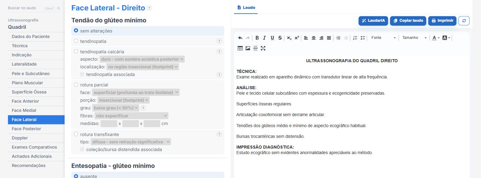
In the hip and joint templates of Laudário:
- Gluteal tendons, bursae and joint recess in their own tabs.
- Laterality and comparison with the contralateral side in the same report.
- Technical limitations in their own tab.
Try it for 15 days free, no credit card required.
The ultrasound reporting platform built by sonologists. We write about reports, classifications and standardization from the real routine of the practice. Who writes and how.
Educational content for healthcare professionals. It does not replace clinical judgment or the guidelines adopted by your service. Editorial policy.