Muscle Injuries on Ultrasound: Strain, Contusion, Hematoma and How to Grade in the Report
When to perform the exam, how to describe the location relative to the myotendinous junction, what to measure, how to grade the injury and the complications that follow-up should look for.
 LaudárioUltrasound reports in practice
This article is part of the guide Musculoskeletal Ultrasound: The Complete Guide, Joint by Joint.
In sports muscle injuries, the sports physician's question is practical: which muscle, exactly where, what size and whether the tendon is involved. This is what estimates the time to return to play. A report that says only "grade II muscle injury" answers little.
When to perform the exam
In the first hours, edema can mask the extent of the injury. An interval of about 24 to 48 hours allows better delineation of the injured area and the hematoma. Serial exams are useful to follow healing.
Strain vs. contusion
- Strain: indirect injury from traction, typical of the myotendinous junction of biarticular muscles (hamstrings, rectus femoris, medial gastrocnemius).
- Contusion: direct trauma, with injury at the point of impact, often deeper, next to the bone, and with hematoma.
What to describe
- The muscle and the region: myotendinous junction, muscle belly or myofascial junction.
- Involvement of the intramuscular tendon or central aponeurosis: this is a poorer prognostic feature, with a longer return to play and more recurrence.
- Extent: craniocaudal length and the approximate proportion of the cross-sectional area involved.
- Hematoma: dimensions and volume.
- Distance from the lesion to a fixed bony landmark, to allow comparison at follow-up.
Grading
The classic ultrasound grading divides injuries into three grades: grade I, minimal change, without evident fiber discontinuity; grade II, partial tear, with discontinuity and hematoma; grade III, complete tear, with retraction of the muscle stump. More recent classifications, such as the British (BAMIC), were developed for MRI and incorporate location and tendon involvement. A report that describes these elements allows any grading system to be applied.
Complications to look for at follow-up
- Myositis ossificans: peripheral calcification that appears weeks after the trauma; recognizing it avoids confusion with a tumor.
- Seroma or posttraumatic cyst: persistent collection.
- Scar fibrosis: hyperechoic, retractile area, associated with recurrence.
- Muscle hernia through the fascia, best seen on contraction.
Common mistakes
- Performing the exam in the first hours and underestimating the injury.
- Grading without stating the muscle and the exact region.
- Not mentioning involvement of the intramuscular tendon.
- Reporting myositis ossificans as an expansile lesion.
See also our article on peripheral nerve ultrasound.
How Laudário helps
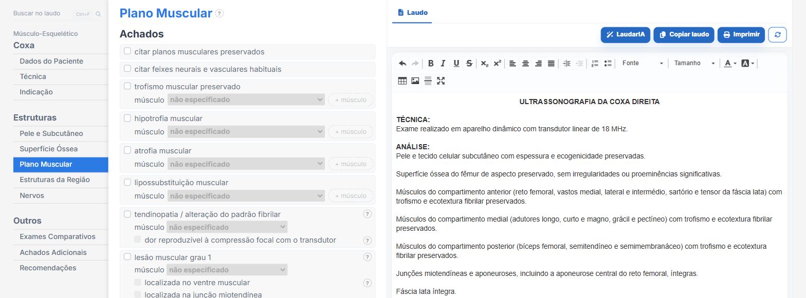
In the soft tissue and musculoskeletal templates of Laudário:
- Numbered lesions with muscle, region, measurements and hematoma.
- Comparison with the previous exam using the same measurements.
- Laterality and recommendations in dedicated fields.
Try it for 15 days free, no credit card.
The ultrasound reporting platform built by sonologists. We write about reports, classifications and standardization from the real routine of the practice. Who writes and how.
Educational content for healthcare professionals. It does not replace clinical judgment or the guidelines adopted by your service. Editorial policy.