Fetal Biometry and Estimated Fetal Weight: Scan Planes, Formulas and Which Chart to Use in the Report
The correct planes for head circumference, abdominal circumference and femur, the error margin of estimated weight, why the pregnancy is not redated by biometry and why the report must state which chart it used.
 LaudárioUltrasound reports in practice
This article is part of the guide Obstetric ultrasound: the three-trimester guide, from dating to Doppler.
Fetal biometry is the most repeated part of the obstetric ultrasound and, for that very reason, the one that accumulates the most small errors. An oblique cut of the head or an abdomen measured at the level of the kidneys changes the estimated weight enough to alter the fetus's classification. A reliable report depends on the right plane and on stating which reference the percentile was calculated with.
The scan planes
- Biparietal diameter and head circumference: transthalamic axial plane, with the thalami and cavum septi pellucidi visible, falx at the midline and no cerebellum in the plane.
- Abdominal circumference: axial plane with the stomach and the intrahepatic portion of the umbilical vein, ideally at the level of the portal sinus; the kidneys should not appear, and the plane should be as circular as possible.
- Femur: the entire ossified diaphysis, with both ends visible and the bone as perpendicular as possible to the beam, without including the epiphysis.
Estimated fetal weight
The Hadlock formulas, which combine head, abdomen and femur, are the most used. Even with good technique, the estimated weight has an error margin on the order of 10 to 15%, larger at the weight extremes. The report should present the value as an estimate.
Dating: the pregnancy is not redated by biometry
Gestational age is defined by the crown-rump length in the first trimester, or by the last menstrual period when concordant. After that, biometry serves to assess growth, not to change the date. A fetus measuring smaller than expected in the third trimester may have growth restriction, and redating hides the problem.
Which chart to use
Hadlock, Intergrowth-21st, the WHO charts and the Fetal Medicine Foundation charts give different percentiles for the same weight. There is no single correct one; what matters is always using the same one in the service and stating in the report which one was used, so that follow-up is comparable.
When the weight departs from the expected
- Below the 10th percentile: small for gestational age; the workup for growth restriction includes Doppler and the Delphi consensus criteria.
- Below the 3rd percentile: isolated criterion for growth restriction.
- Above the 90th percentile: large for gestational age; correlation with gestational diabetes and assessment of the amniotic fluid.
Common errors
- Head circumference with the cerebellum in the plane, or abdominal circumference with the kidneys.
- Redating the pregnancy in the third trimester by biometry.
- Percentile without the reference chart.
- Presenting the estimated weight as an exact measurement.
See also the texts on fetal growth restriction and obstetric Doppler.
How Laudário helps
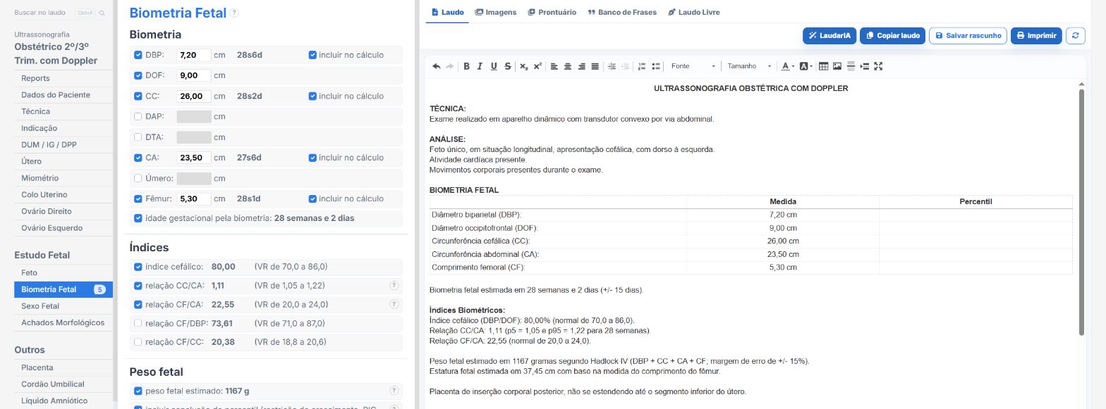
In the obstetrics templates of Laudário:
- Estimated fetal weight calculated from the measurements, with percentile.
- Reference chart chosen by the physician and cited in the report.
- Gestational age by the reference date, without redating by biometry.
Try it for 15 days free, no credit card.
The ultrasound reporting platform built by sonologists. We write about reports, classifications and standardization from the real routine of the practice. Who writes and how.
Educational content for healthcare professionals. It does not replace clinical judgment or the guidelines adopted by your service. Editorial policy.