Intussusception in children: the target sign, what to measure and what to report
How to look for ileocolic intussusception on ultrasound, the target and pseudokidney signs, how to tell it from transient small bowel intussusception and which findings change the reduction decision.
 LaudárioUltrasound reports in practice
Intussusception is the most common cause of bowel obstruction between 6 months and 3 years of age. Ultrasound has very high sensitivity, uses no radiation and, in many services, also guides hydrostatic reduction. The report needs to confirm or exclude the diagnosis and provide the findings that influence the decision between reduction and surgery.
Technique
- High-frequency linear transducer, sweeping the course of the colon: right iliac fossa, right flank, right upper quadrant, epigastrium and left colon.
- Ileocolic intussusception is usually in the right upper quadrant, near the liver.
- Also assess the right iliac fossa to locate the cecum.
The signs
- Target sign (or doughnut sign) on the transverse view: concentric rings formed by the walls of the bowel loops, usually 3 to 5 cm in diameter.
- Pseudokidney sign on the longitudinal view.
- Mesenteric fat and lymph nodes dragged into the intussusception.
Ileocolic or transient small bowel
Intussusceptions between small bowel loops that are small (usually less than 2.5 cm in diameter), short, with preserved peristalsis and no obstruction, are often transient and resolve on their own. The report should differentiate them from ileocolic intussusception, because management is different: observation and reassessment, not reduction.
Findings that change management
- Doppler flow in the walls of the intussusceptum: its presence is favorable; its absence raises suspicion of vascular compromise.
- Trapped fluid between the layers, associated with a lower chance of reduction success and with ischemia.
- Free fluid with debris or signs of perforation.
- Pathologic lead point (Meckel diverticulum, polyp, duplication, lymphoma), more likely in children outside the typical age range.
- Signs of obstruction, with distended small bowel loops.
Peritonitis, perforation and instability are contraindications to reduction and indicate surgery.
What the report should include
- Location and type (ileocolic, ileoileal).
- Diameter and, if possible, length.
- Wall Doppler, trapped fluid and free fluid.
- Lead point, when identified.
- Signs of obstruction.
- If guided reduction was performed: technique, result and confirmation of passage into the terminal ileum.
Common mistakes
- Not sweeping the entire course of the colon and looking only in the right iliac fossa.
- Reporting a transient small bowel intussusception as ileocolic.
- Omitting the Doppler and trapped fluid.
- Not looking for a lead point in a child outside the typical age range.
- After reduction, not confirming resolution and passage into the ileum.
See also our text on ultrasound in appendicitis.
How Laudário helps
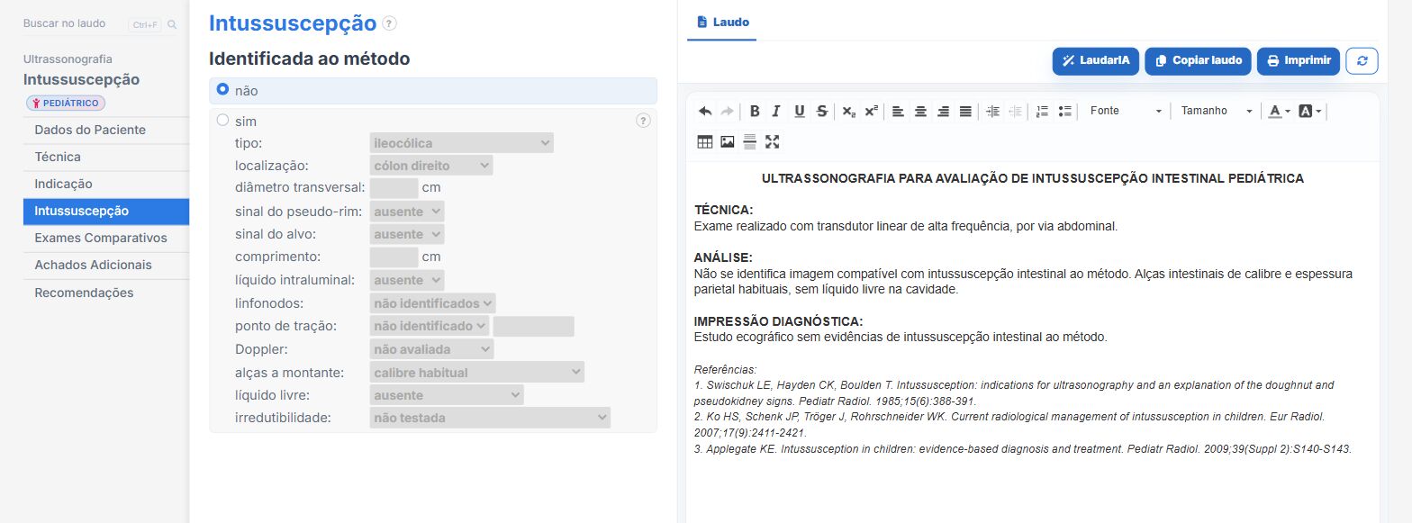
In the pediatric bowel intussusception template of Laudário:
- Location, type and measurements in their own fields.
- Doppler, trapped fluid, free fluid and lead point as options.
- Conclusion and recommendations assembled from the findings.
Try it for 15 days free, no credit card required.
The ultrasound reporting platform built by sonologists. We write about reports, classifications and standardization from the real routine of the practice. Who writes and how.
Educational content for healthcare professionals. It does not replace clinical judgment or the guidelines adopted by your service. Editorial policy.