Infant hip ultrasound: how to structure the report when assessing developmental dysplasia
Infant hip ultrasound is the main imaging exam for early diagnosis of developmental dysplasia of the hip (DDH). See how to structure the report using the Graf method, with clear classification, measured angles and guided management, all standardized and auditable.
 LaudárioUltrasound reports in practice
Infant hip ultrasound is the main imaging study for screening and diagnosing developmental dysplasia of the hip (DDH) in the first months of life. Because it is performed in a window when the ossification centers are still predominantly cartilaginous, it can assess acetabular morphology and the stability of the femoroacetabular relationship with sensitivity far superior to radiography. The trade-off is a heavy dependence on standardized technique and on a clear report: small variations in positioning or description can change management, from simple follow-up to the use of a Pavlik harness. The report therefore needs to be objective, reproducible and directly guide the pediatric orthopedist.
Why standardize the infant hip ultrasound report
DDH is one of the main causes of limping and early hip osteoarthritis in adults, and ultrasound screening in the newborn and young infant is the most effective way to identify it before it causes permanent deformity. In many practices, the study is performed in every newborn with a risk factor (breech presentation, positive family history, female sex, obvious ligamentous laxity, oligohydramnios) or physical exam findings (positive Ortolani and Barlow maneuvers, skin fold asymmetry).
Without standardization, it is common for the report to state only “hips without abnormalities” or “preserved morphology”, without citing the angles measured, the Graf type assigned or the technique used. This compromises comparison with future studies, makes auditing the service harder and, above all, delays the orthopedist's decision-making. A well-structured report describes the technique, provides the measurements, classifies according to Graf and concludes with explicit clinical guidance.
Ideal window for the exam and data that should appear in the identification
Infant hip ultrasound is most informative between the 4th and 12th week of life. Before that, physiological neonatal laxity can generate false positives; after the 4th month, progressive ossification of the femoral head begins to shadow the acetabulum and reduce the accuracy of the Graf method, at which point the AP pelvis radiograph becomes the study of choice. The report should always record the exact age in weeks or months, making clear whether the window still allows complete morphological analysis.
Before the description itself, the report needs to give context to the study: clinical indication (screening, risk factor or physical exam finding), patient age, position used (lateral decubitus is the standard for the Graf technique), degree of cooperation and quality of the window. When the study is serial, it is worth citing the date and classification of the prior study, recording whether there was improvement, stability or worsening.
The Graf technique and what the report needs to describe
The Graf technique is the most established method for morphological assessment of the infant hip. It requires a standard coronal plane in which three structures must be visible simultaneously: the lower limb of the ilium in the acetabular fossa, the acetabular labrum and the straight line of the ilium. Without this triad, the plane is not valid and any measurement will be unreliable. This point should be made clear in the report, avoiding classifying hips from images outside the standard.
In the correct plane, two reference lines are drawn on which the method rests:
- Alpha angle (α): formed between the baseline (parallel to the iliac line) and the bony roof line of the acetabulum. It reflects the maturity of the bony component of the acetabulum. The larger the angle, the more mature the hip.
- Beta angle (β): formed between the baseline and the cartilaginous roof line (acetabular labrum). It reflects the cartilaginous coverage. In unstable or dislocated hips, beta increases progressively.
Both angles should be measured for each hip separately and recorded in the report. Simply assigning a Graf type without recording the numeric values makes the study less auditable and complicates comparison over time.
Graf classification: what each type means
The main types of the Graf classification, with their reference values most commonly used in clinical practice, should be clearly described in the report:
- Type I (mature): α ≥ 60° and β ≤ 55°. Hip with normal morphology for age. Usual management: discharge from screening.
- Type IIa (physiologically immature): α between 50° and 59° in a child younger than 12 weeks. Reflects immaturity consistent with age. Usual management: reassessment around the 12th week.
- Type IIb (immature after 12 weeks): same α range, but in a child older than 12 weeks. This already constitutes mild dysplasia and calls for orthopedic evaluation.
- Type IIc (critical): α between 43° and 49°, with β < 77°. Dysplastic hip, still centered, but at risk of decompensation. Requires close orthopedic follow-up.
- Type D (decentering): α between 43° and 49°, with β ≥ 77°. Hip on the verge of dislocation, with usual indication for treatment.
- Type III (dislocated): α < 43°. Femoral head displaced laterally, with the acetabular labrum still preserved (IIIa) or already showing echogenic changes (IIIb).
- Type IV (dislocated with inverted labrum): femoral head outside the acetabulum, with the acetabular labrum interposed. Most severe form, with frequent surgical indication.
The classification should be given separately for each side, even when both are symmetric. In discordant cases, the report must make clear the type of each hip, avoiding generic descriptions such as “symmetric, mature hips” when only one was adequately assessed.
Dynamic assessment: stability and stress tests
Beyond morphology, infant hip ultrasound allows assessment of joint stability through dynamic maneuvers, notably the modified Barlow maneuver performed under ultrasound visualization. The report should record whether the hip remained centered, whether there was reproducible subluxation (partial displacement of the femoral head relative to the acetabulum) or frank dislocation during the maneuver.
This information is particularly relevant in hips classified as IIc and D: clinical stability on dynamic ultrasound helps the orthopedist decide between close observation and starting a harness. Describing stability objectively, without vague terms such as “apparently stable”, increases the value of the report.
Conclusion and clinical guidance
The conclusion of the report should bring, in a few lines, three pieces of information that the pediatric orthopedist looks for immediately:
- The Graf type assigned to each hip, with the corresponding α and β angles.
- The dynamic status of the hip (stable, subluxable or dislocated).
- The suggested follow-up, adjusted to the age group and the classification found: reassessment in 4 to 6 weeks for IIa, referral to the orthopedist for type IIc and above, discharge for type I.
It is important to remember that hip ultrasound does not replace the orthopedist's clinical evaluation. Even in reports classified as Type I, it is prudent to emphasize that if suspicious clinical findings persist (skin fold asymmetry, limited abduction, apparent shortening), imaging reassessment should be repeated.
Findings that deserve emphasis in the conclusion
- Asymmetry between the hips, even if both are within normal limits, especially in patients with a risk factor.
- Hip classified as IIa in a child close to 12 weeks, a window in which the natural evolution to IIb may be underway.
- Progressive increase of the beta angle on serial studies, an early sign of instability.
- Discordance between the measured angles and the clinical finding (for example, Graf I with a positive Ortolani sign): it is worth recording the observation and suggesting reassessment.
- Suboptimal acoustic window due to poor cooperation or advanced ossification of the femoral head: report it transparently, avoiding underestimating the limitation of the method.
Precautions to avoid ambiguous reports
- Always record the α and β angles in degrees, not just the Graf type.
- Describe the side assessed explicitly (right and left), avoiding collective terms such as “hips”.
- Document the quality of the coronal plane and the presence of the three reference structures.
- In patients beyond the ideal window, state the technical limitation and consider recommending an AP pelvis radiograph.
- Avoid absolute conclusive phrases (“normal”, “without abnormalities”) when there are borderline findings: prefer describing the values and giving context for the age group.
How Laudário helps structure the infant hip ultrasound report
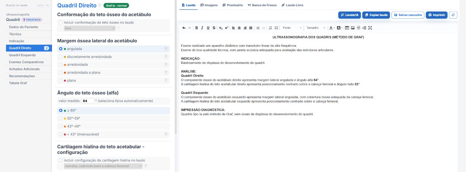
Laudário was developed to speed up report typing without losing the clinical depth that studies such as infant hip ultrasound demand. Within the system, the Infant Hip template already comes with the complete structure of the Graf method, with ready-made fields to describe the technique, record the α and β angles, assign the classification type for each side and automatically generate the standardized conclusion.
In practice, this means:
- A ready-made structure to describe the coronal plane, the reference triad and the stability maneuvers, with standardized phrases adjustable to your reporting style.
- Automatic Graf classification from the angles entered, eliminating classification errors and discrepancies between the body of the report and the conclusion.
- Integration with other pediatric and musculoskeletal templates, favoring consistency across serial reports of the same patient.
- Professional printing with a QR Code and a digital version of the report, adding traceability and more perceived value when delivering it to the patient, the pediatrician and the orthopedist.
- Support from LaudarIA, the integrated artificial intelligence, to help build phrases, conclusions and fine adjustments to the text, especially useful in borderline cases where the wording makes a difference to the clarity of the report.
The result is a standardized infant hip report that is quick to produce and, above all, more useful to the pediatric orthopedist who will receive the document. In a high-volume ultrasound practice, saving seconds per report and standardizing clinical communication means less rework, more productivity and higher quality in delivery.
If you do not know it yet, it is worth trying: Laudário offers a 15-day free trial, no credit card required, with immediate access to all exam templates, including infant hip and the other protocols of pediatric, musculoskeletal and general ultrasound practice.
The ultrasound reporting platform built by sonologists. We write about reports, classifications and standardization from the real routine of the practice. Who writes and how.
Educational content for healthcare professionals. It does not replace clinical judgment or the guidelines adopted by your service. Editorial policy.