Cranial ultrasound: how to structure the report in the neurologic evaluation of the newborn
Cranial ultrasound through the fontanelle is the first-line study for neurologic evaluation of the newborn. See how to structure the report consistently: indications, technique, peri-intraventricular hemorrhage grading (Papile), leukomalacia (De Vries), ventriculomegaly and the most frequent errors.
 LaudárioUltrasound reports in practice
Transfontanellar (cranial) ultrasound is today the first-choice imaging study for the neurologic evaluation of the newborn, especially premature infants admitted to the NICU. Available at the bedside, with no need for sedation and no ionizing radiation, it allows early identification of germinal matrix hemorrhage, periventricular leukomalacia, hydrocephalus and congenital malformations that directly change clinical management.
Although it is a relatively simple study from a technical standpoint, the transfontanellar report is often a source of variability between practitioners: sometimes too brief, sometimes too descriptive, sometimes without standardized grading of the findings. In this post, we outline what a consistent, objective transfontanellar ultrasound report that is useful to the neonatologist managing the patient must include.
When transfontanellar ultrasound is indicated
The classic indication is routine evaluation of the premature newborn, especially below 32 weeks and/or with birth weight under 1,500 g. In this population, the American Academy of Neurology recommends routine screening between the 7th and 14th day of life and a new evaluation between 36 and 40 weeks of postmenstrual age, precisely because of the increased risk of peri-intraventricular hemorrhage and white matter injury.
Besides screening of the premature infant, the study is requested in term newborns with clinical signs suggesting neurologic involvement: seizures, marked hypotonia, abnormal head circumference, bulging fontanelle, suspected congenital infection (TORCH), neonatal sepsis, perinatal asphyxia or after therapeutic cooling. In any scenario, the open fontanelle window makes the method especially attractive: as long as the anterior fontanelle remains patent (usually up to 12 to 18 months), the evaluation can be repeated as many times as necessary, at low cost and with no risk to the patient.
Recommended report structure
A good transfontanellar report follows the same logic as any structured ultrasound report: clear identification of the study, technique used, systematic description of the structures evaluated and an objective conclusion. The difference is that, in the neonate, small findings can have great clinical impact, so standardization is what prevents shallow or ambiguous reports.
1. Technique and acoustic windows
Describe which transducer was used (microconvex or sector, 5 to 10 MHz, is the most common) and which acoustic windows were employed. The anterior fontanelle is the main access, with coronal and sagittal/parasagittal sweeps. When available, also record the use of the mastoid window (excellent for the posterior fossa and cerebellum) and the posterior window, which are essential for a complete evaluation.
2. Structures that must be described
- Cerebral parenchyma: echogenicity, differentiation between white and gray matter, symmetry between hemispheres.
- Ventricular system: lateral ventricles (body, atrium, frontal, occipital and temporal horns), third and fourth ventricles. Measure the ventricular atrium on the coronal view (reference value below 10 mm).
- Extra-axial CSF spaces: subarachnoid space, cisterna magna, interhemispheric fissure.
- Midline structures: corpus callosum, cavum septi pellucidi, choroid plexuses.
- Basal ganglia and thalami: echogenicity and symmetry.
- Posterior fossa: vermis and cerebellar hemispheres, fourth ventricle, brainstem (preferably through the mastoid window).
3. Grading of peri-intraventricular hemorrhage
This is the most frequent finding, and the one that most calls for standardization. The Papile classification, still widely used in practice, divides PIVH into four grades:
- Grade I: hemorrhage confined to the germinal matrix (caudothalamic groove), without extension into the ventricle.
- Grade II: intraventricular hemorrhage without ventricular dilatation.
- Grade III: intraventricular hemorrhage with ventricular dilatation.
- Grade IV: hemorrhagic infarction of the adjacent periventricular parenchyma (currently classified separately as venous hemorrhagic infarction).
Always report the side involved, the extent, the presence of intraventricular clot and the evolutionary appearance (acute hyperechoic hemorrhage, organizing, or in a retraction phase with cystic appearance). When possible, compare with prior studies: temporal evolution is an essential part of the neonatal report.
4. Periventricular leukomalacia and white matter injury
Periventricular leukomalacia (PVL) is the leading cause of cerebral palsy in premature infants and deserves careful description. Characterize the persistent periventricular hyperechogenicity, the presence of cysts (cystic PVL) and their location: frontal, peritrigonal or occipital. The De Vries grading is useful:
- Grade I: periventricular hyperechogenicity persisting for more than 7 days, without cysts.
- Grade II: hyperechogenicity that evolves into small localized frontoparietal cysts.
- Grade III: extensive periventricular occipitoparietal cysts.
- Grade IV: cysts extending into the subcortical white matter.
5. Hydrocephalus and ventriculomegaly
Whenever there is ventricular enlargement, measure the ventricular atrium on the coronal view and, ideally, the Levene index (distance between the midline and the lateral edge of the ventricle). Differentiate ventriculomegaly (dilatation without increased CSF pressure) from hydrocephalus (with signs of hypertension), state whether it is unilateral or bilateral, symmetric or asymmetric, and whether the third and fourth ventricles are involved. In post-hemorrhagic cases, serial follow-up is essential.
6. Other relevant findings
- Choroid plexus cysts: characterize size and laterality, and describe as an isolated finding when applicable.
- Subependymal cyst: frequent in congenital infections (CMV) and after previous hemorrhage.
- Calcifications: suggest congenital infection; describe location (periventricular in CMV, scattered in toxoplasmosis).
- Malformations: agenesis of the corpus callosum, holoprosencephaly, Dandy-Walker: complete description and indication for complementary MRI when appropriate.
- Hypoxic-ischemic encephalopathy: diffuse cerebral edema, loss of white-gray matter differentiation, hyperechogenicity in the basal ganglia and thalami.
Report conclusion: objective and useful to the clinician
The conclusion should be direct, summarize the main findings and their classification (Papile and De Vries grades, ventricular measurement), and, when pertinent, suggest follow-up (repeat study in 7 to 14 days, complementary MRI, pediatric neurology evaluation). Avoid repeating the entire description: the neonatologist needs a clear synthesis to make a decision.
Frequent errors in the transfontanellar report
- Not grading the peri-intraventricular hemorrhage, or using inconsistent terminology between reports from the same institution.
- Forgetting to measure the ventricular atrium on coronal views, especially in cases with suspected dilatation.
- Omitting the mastoid window: the posterior fossa is underdiagnosed when evaluated only through the anterior fontanelle.
- Describing findings without comparing with prior studies in patients under serial follow-up.
- A generic conclusion such as “study without significant abnormalities” without detailing what was evaluated.
How Laudário helps standardize the transfontanellar report
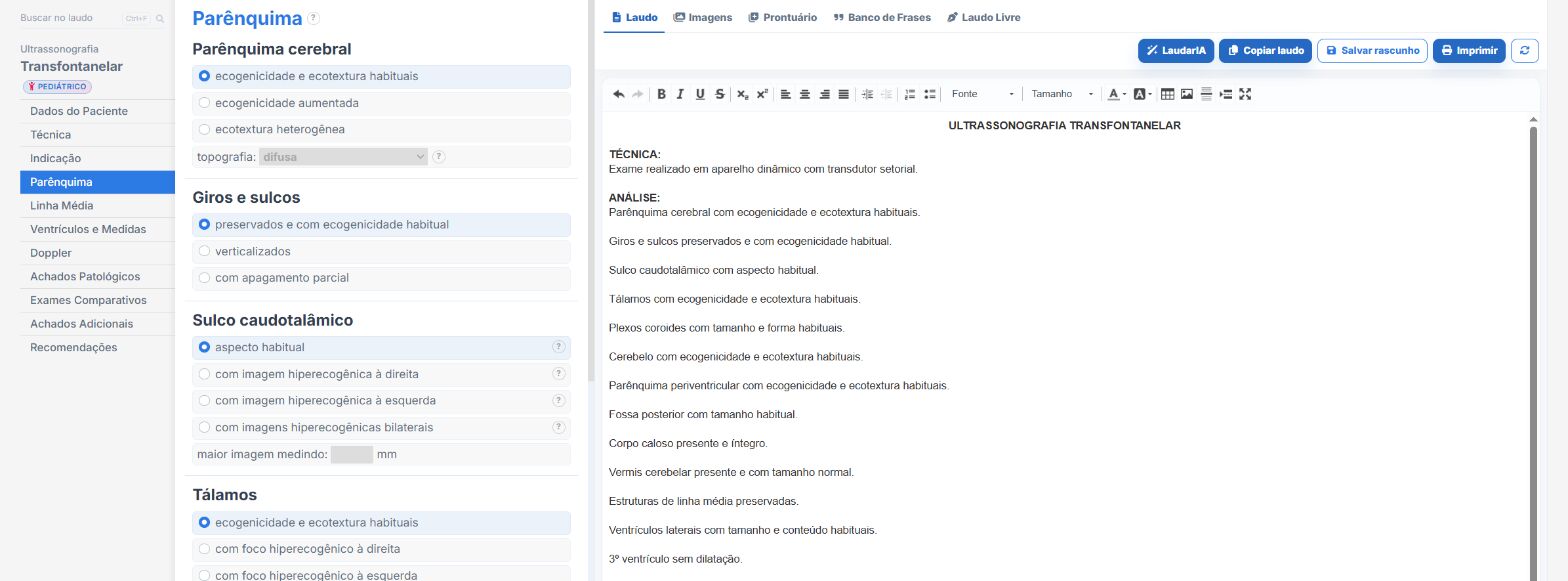
Many of the difficulties above stem from something simple: typing a neonatal report from scratch, in the middle of the NICU routine, under time pressure and with multiple patients. That is where variations in wording, findings that escape the description and conclusions that do not follow the standard grading in the literature come in.
Laudário was designed for exactly this scenario. With structured templates for each study, transfontanellar ultrasound, like the other 58 exams available on the platform, already comes with the sequence of structures to describe, ready-made phrases for the main findings and classifications integrated into the typing flow. You gain speed without losing standardization, keep consistency across the team's reports and reduce the risk of forgetting an important item in the rush of a shift.
For clinics and imaging services that care for neonatology, this means more predictable reports, faster to produce and with the credibility the neonatologist needs to manage the patient safely. Want to see how it works in practice? Try Laudário free for 15 days: no credit card, immediate access and cancellation at any time.
The ultrasound reporting platform built by sonologists. We write about reports, classifications and standardization from the real routine of the practice. Who writes and how.
Educational content for healthcare professionals. It does not replace clinical judgment or the guidelines adopted by your service. Editorial policy.