Hashimoto Thyroiditis on Ultrasound: Parenchyma, Pseudonodules and the Differential with Graves
Diffuse hypoechogenicity, micronodular pattern and fibrous septa, the pseudonodules that should not receive a TI-RADS category, the Doppler that separates Graves from destructive thyroiditis, and subacute thyroiditis.
 LaudárioUltrasound reports in practice
Not every thyroid ultrasound is about nodules. A large share of requests come from patients with hypothyroidism, positive antibodies or goiter, and the question is about the parenchyma: are there signs of thyroiditis? In most cases, the answer is Hashimoto thyroiditis, the most common cause of hypothyroidism in iodine-sufficient regions.
This text shows the parenchymal findings in thyroiditis, how to tell them apart and the care the report needs to take with "pseudonodules", which lead to unnecessary biopsies.
The normal parenchyma
The normal thyroid is homogeneous and more echogenic than the pretracheal strap muscles. The report records the measurements of the lobes and isthmus and the volume of each lobe (three diameters multiplied by 0.523), with the sum. The total volume in adults is generally below 18 mL in women and 25 mL in men, with regional variation.
Hashimoto thyroiditis
Chronic autoimmune thyroiditis produces changes that reflect lymphocytic infiltration and, over time, fibrosis:
- Diffuse hypoechogenicity, sometimes equal to or lower than that of the muscles: this is the most characteristic finding.
- Heterogeneity with 1 to 6 mm hypoechoic micronodules surrounded by echogenic septa, the "micronodular" pattern.
- Echogenic fibrous septa, which give a pseudolobulated appearance.
- Volume increased in the early stages (goiter) and reduced in the late stages (atrophy).
- Variable vascularity: normal, increased or reduced.
- Reactive lymph nodes at level VI, below the isthmus, frequent and benign.
Ultrasound suggests thyroiditis, but the diagnosis is clinical and laboratory-based (anti-TPO and antithyroglobulin antibodies). The report should describe the parenchyma and conclude "sonographic appearance consistent with diffuse thyroid disease", recommending laboratory correlation, and not state Hashimoto without the antibodies.
Pseudonodules
In thyroiditis, areas of better-preserved or more heavily infiltrated parenchyma can look like nodules. The most typical are the hyperechoic or "white" nodules, which in thyroiditis are almost always benign (regenerative hyperplasia). Pseudonodules are recognized by:
- Not being consistently seen in two orthogonal planes.
- Margins that blend into the surrounding parenchyma.
- Not displacing the vessels.
Applying TI-RADS to a pseudonodule leads to unnecessary biopsies. On the other hand, a true nodule in thyroiditis should be classified like any other. It is worth remembering that patients with Hashimoto have an increased risk of thyroid lymphoma: rapid enlargement of the gland or a markedly hypoechoic mass deserves investigation.
Graves disease
This is the main differential for the hyperthyroid phase of Hashimoto (hashitoxicosis). In Graves disease:
- Enlarged, hypoechoic gland.
- Intense, diffuse hypervascularity on Doppler, the so-called "thyroid inferno".
- Elevated velocities in the inferior thyroid arteries.
Doppler helps separate hyperthyroidism from overproduction (Graves, with increased flow) from thyrotoxicosis from destruction (subacute and painless thyroiditis, with reduced flow), a distinction that changes treatment.
Subacute (De Quervain) thyroiditis
Neck pain, fever and thyrotoxicosis after a viral illness. Ultrasound shows ill-defined hypoechoic areas, geographic, with reduced vascularity, sometimes migrating from one lobe to the other. These areas mimic suspicious nodules: the clinical context and the course (regression over weeks to months) avoid biopsy. When in doubt, the report recommends sonographic follow-up, not TI-RADS.
How to structure the report
- Dimensions and volume of each lobe, isthmus and total volume.
- Echogenicity (normal, reduced, markedly reduced) and texture (homogeneous, heterogeneous, micronodular).
- Vascularity on Doppler.
- True nodules with TI-RADS; pseudonodules described as such.
- Cervical lymph nodes.
- Descriptive diagnostic impression, with a recommendation for laboratory correlation.
For nodules, see our article on thyroid and TI-RADS.
Common mistakes
- Stating Hashimoto without antibodies.
- Classifying a pseudonodule by TI-RADS.
- Omitting the volume.
- Not using Doppler in thyrotoxicosis.
- Recommending biopsy of a subacute thyroiditis area.
How Laudário helps
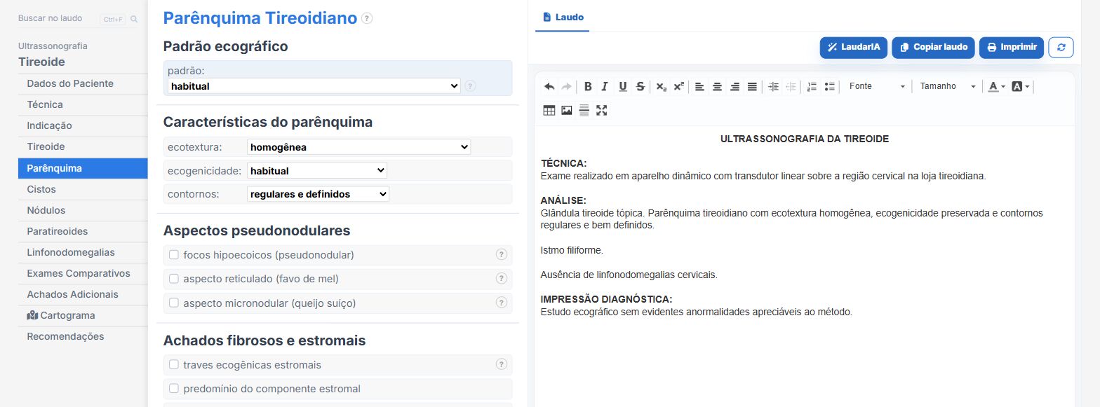
In the thyroid templates of Laudário, with and without Doppler:
- Volume of each lobe and total calculated automatically.
- Diffuse thyroid disease parenchymal patterns as options.
- Nodules with calculated TI-RADS and a cartogram.
- Doppler vascularity and cervical lymph nodes.
Try it for 15 days free, no credit card.
The ultrasound reporting platform built by sonologists. We write about reports, classifications and standardization from the real routine of the practice. Who writes and how.
Educational content for healthcare professionals. It does not replace clinical judgment or the guidelines adopted by your service. Editorial policy.