Neck ultrasound: how to structure the report for cervical lymph nodes and masses
Neck ultrasound is one of the most requested imaging studies in outpatient practice and serves as the entry point for investigating lymphadenopathy, palpable masses and abnormalities of the superficial glands of the neck. Its great advantage is excellent resolution for superficial structures...
 LaudárioUltrasound reports in practice
Neck ultrasound is one of the most requested imaging studies in outpatient practice and serves as the entry point for investigating lymphadenopathy, palpable masses and abnormalities of the superficial glands of the neck. Its great advantage is excellent resolution for superficial structures combined with color Doppler, which allows the vascular pattern of lymph nodes and nodules to be characterized in a level of detail that other methods only reach with contrast. The trade-off is a complex regional anatomy (multiple nodal levels, salivary glands, thyroid, neck vessels and muscular structures), which makes the report particularly vulnerable to generic descriptions. A well-structured report is what separates a “nonspecific cervical lymphadenopathy” from useful clinical guidance that correctly directs the otolaryngologist, oncologist, pediatrician or attending physician.
Why standardize the neck ultrasound report
Neck ultrasound covers a very broad diagnostic spectrum: investigation of reactive and neoplastic lymphadenopathy, evaluation of congenital masses (thyroglossal duct cysts, branchial cysts), study of the salivary glands (sialolithiasis, sialadenitis, parotid nodules), head and neck oncologic follow-up, post-thyroidectomy surveillance, and investigation of muscular and vascular masses. Without standardization, the same study can receive completely different reports depending on the operator: some describe only the dominant finding, others list structures with no clinical hierarchy.
A well-structured neck report describes the anatomic region by nodal level, characterizes each relevant finding with objective criteria (dimensions, shape, hilum, echogenicity, vascularity) and concludes by guiding the clinical hypothesis and management. This standardization is especially important in oncology patients, in whom serial comparison between studies is a central part of follow-up.
Indications and data that should appear in the identification
The neck ultrasound report should state the clinical indication right at the start: investigation of a palpable mass, lymphadenopathy found on physical examination, local symptoms (dysphagia, odynophagia, hoarseness), oncologic follow-up, postoperative surveillance, or screening in a patient with a risk factor. When the study is serial, as is common in oncology patients, it is worth recording the date of the previous study and explicitly stating whether the comparison is favorable, stable or unfavorable.
It is also important to record the target region of the study: complete neck (all levels), neck focused on a specific level, lymph nodes only, or neck including the salivary glands and/or thyroid. Defining this scope prevents the report from being read as incomplete when the focus of the study was in fact specific.
Anatomy by levels: the basis of the neck report
The nodal level classification, originally proposed for staging head and neck tumors and widely adopted in sonographic practice, is the natural backbone of the neck report. Describing findings by level standardizes communication with the surgeon and the oncologist, avoiding ambiguous terms such as “lymph node in the right submandibular region” without further topographic detail.
- Level I: submental (IA) and submandibular (IB). Drains the lips, tip of the tongue, floor of the mouth and submandibular glands.
- Level II: upper jugular, from the skull base to the hyoid bone. Drains the nasopharynx, oropharynx, parotid and posterior oral cavity.
- Level III: mid jugular, from the hyoid to the lower border of the cricoid cartilage. Drains the hypopharynx and larynx.
- Level IV: lower jugular, from the cricoid to the clavicle. Drains the thyroid, cervical esophagus and upper thoracic structures.
- Level V: posterior triangle, from the mastoid to the clavicle, behind the sternocleidomastoid. Drains the nasopharynx, posterior scalp and thyroid.
- Level VI: central compartment, between the carotids, from the hyoid to the sternal notch. It is the main drainage chain of the thyroid.
- Level VII: superior mediastinum, only partially accessible to ultrasound in thin patients.
For each level evaluated, the report should state whether lymph nodes are visualized, describe the findings when relevant and record laterality (right, left or bilateral). The simple mention of “unremarkable cervical chains” should be replaced by a description that explicitly covers the levels studied.
How to characterize a lymph node in the report
The description of a cervical lymph node needs to go beyond its dimensions. The sonographic criteria established in the literature allow suspicion to be stratified between reactive nodes and nodes suspicious for neoplastic or specific inflammatory involvement. Each relevant lymph node should be described in terms of:
- Location: nodal level and laterality.
- Dimensions: record the longest longitudinal axis (L) and the longest transverse axis (T) in millimeters. A short axis above 10 mm at levels I to V (and above 8 mm at level II) is classically considered borderline.
- L/T ratio (Solbiati index): benign nodes tend to keep an oval shape (L/T > 2). Round nodes (L/T < 2) are suspicious.
- Echogenic hilum: its preservation is one of the most reliable signs of benignity. Loss of the hilum, replaced by a diffuse hypoechoic pattern, raises suspicion.
- Cortical echogenicity: describe focal cortical thickening, a heterogeneous pattern, areas of intranodal necrosis or microcalcifications (the latter a classic finding of papillary thyroid carcinoma metastasis).
- Margins: well defined (more common in benign nodes) or poorly defined/blurred (raising the possibility of extracapsular extension in a neoplastic context).
- Vascularity on color Doppler: central hilar pattern (typical of benignity), peripheral/capsular or mixed disorganized pattern (more associated with malignancy).
The combination of these criteria matters more than any isolated parameter. A 14 mm lymph node with an oval shape, preserved hilum and central hilar vascularity, in a young patient with a recent infection, has a typical reactive character. A round 9 mm node with no hilum, with microcalcifications and peripheral vascularity, in a patient with a previous thyroidectomy for papillary carcinoma, requires a detailed description and an explicit recommendation for further evaluation.
Non-nodal masses: what must not be mistaken
Neck ultrasound frequently identifies findings that are not lymph nodes but must be correctly categorized in the report. Mistaking a thyroglossal duct cyst for a cystic lymph node, or a nodule in the tail of the parotid for a level II lymphadenopathy, leads to inappropriate management.
- Thyroglossal duct cyst: anechoic or hypoechoic lesion, usually midline or paramedian, between the hyoid and the thyroid, moving with swallowing.
- Branchial cyst: usually lateral, anterior to the sternocleidomastoid, at level II or III, often with proteinaceous content.
- Salivary gland nodules: should be described in terms of the gland involved (parotid, submandibular, sublingual), lobar location (superficial or deep in the parotid), dimensions, echogenicity, margins and vascularity.
- Sialolithiasis: hyperechoic focus with posterior acoustic shadowing along the ductal course, often accompanied by upstream ductal dilation.
- Thyroid nodules found during the neck study: should be described using TI-RADS criteria, even if the main focus of the study is nodal.
- Vascular and neurogenic structures: the carotid bulb, paragangliomas, schwannomas and neuromas require careful identification before being labeled as lymphadenopathy.
Conclusion and clinical guidance
The conclusion of the neck report should synthesize, in a few lines, three essential pieces of information for the referring physician: the presence or absence of lymphadenopathy with suspicious features, the location and stratification of the dominant finding, and the suggested management, whether clinical follow-up, comparison with a future study, specialist evaluation or an indication for biopsy/FNA.
In oncology patients, the conclusion must make the serial comparison explicit: dimensional stability, reduction relative to the previous study, or dimensional/numerical progression. Vague phrases such as “cervical lymph nodes without significant abnormalities” are of little use in this context and should be replaced by an objective comparison that cites the previous study.
Findings that deserve emphasis in the conclusion
- A lymph node with loss of the hilum associated with a round shape (L/T < 2), especially in an oncologic context or in a patient with a previous head and neck neoplasm.
- Intranodal microcalcifications or areas of cystic degeneration, particularly in the follow-up of papillary thyroid carcinoma.
- Peripheral or mixed disorganized vascularity on color Doppler.
- Confluent lymph nodes or nodes with a mass-like appearance, raising the possibility of a nodal conglomerate.
- Incidental extranodal findings, such as thyroid nodules, congenital cysts or salivary gland masses.
- Dimensional or numerical progression on serial studies, even if each individual lymph node remains below the classic threshold of suspicion.
Precautions to avoid ambiguous reports
- Always describe findings by nodal level and laterality, avoiding collective terms such as “cervical chains”.
- Record dimensions in millimeters and the L/T ratio, instead of qualitative descriptions such as “mild lymphadenopathy”.
- Characterize the vascular pattern on color Doppler: the absence of this description substantially reduces the diagnostic value of the study.
- Clearly differentiate lymph nodes from other cervical structures (congenital cysts, salivary nodules, vessels), avoiding generic labels.
- In oncology patients, provide an explicit serial comparison; simply repeating descriptions from the previous study is not enough.
- Reserve absolute conclusions (“unremarkable”) for truly normal studies; for borderline findings, prefer to describe them transparently and suggest reassessment.
How Laudário helps structure the neck ultrasound report
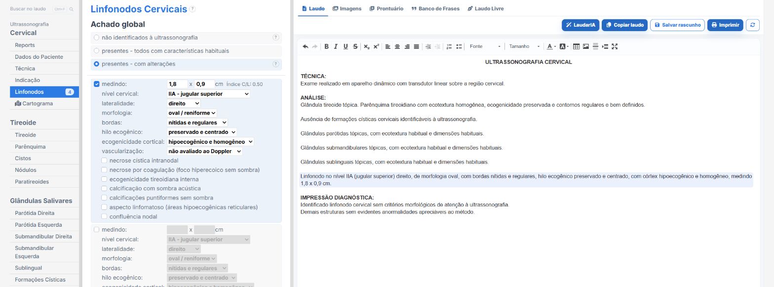
Laudário was developed to speed up report typing without losing the clinical depth that studies such as neck ultrasound demand. Within the system, the Cervical and Cervical (lymph nodes only) templates already come with the complete structure by nodal level, with ready-made fields to describe laterality, dimensions, L/T ratio, hilum, echogenicity, margins and Doppler vascularity, exactly the criteria that separate an objective report from a generic description.
In practice, this means:
- A ready structure to describe each nodal level, with standardized phrases adjustable to your reporting style.
- Dedicated fields for dimensions and the L/T ratio, preventing important criteria from being forgotten during fast typing.
- Integration with the Thyroid and Salivary Glands templates, making combined reports easier when the neck study identifies relevant incidental findings.
- A standardized conclusion that can be adjusted to reflect both normal studies and oncologic follow-ups with serial comparison.
- Professional printing with a QR Code and a digital version of the report, adding traceability and more perceived value when delivering it to the patient and the referring physician.
- Support from LaudarIA, the integrated artificial intelligence, to help build phrases, conclusions and fine adjustments to the text, particularly useful in combined descriptions in which lymph nodes, salivary nodules and thyroid nodules coexist.
The result is a standardized neck report that is quick to produce and, above all, more useful to the physician who requested the study. In a high-volume ultrasound practice, standardizing communication by nodal level and keeping the description of lymph nodes always complete means less rework, greater diagnostic confidence and more added value in every study delivered.
If you are not yet familiar with it, it is worth trying: Laudário offers a 15-day free trial, with no credit card, with immediate access to all study templates, including the neck, the neck lymph nodes only and the other head and neck protocols of the routine.
The ultrasound reporting platform built by sonologists. We write about reports, classifications and standardization from the real routine of the practice. Who writes and how.
Educational content for healthcare professionals. It does not replace clinical judgment or the guidelines adopted by your service. Editorial policy.