Salivary gland ultrasound: parotid, submandibular, stones, Sjögren and nodules
The lobe of the parotid nodule relative to the retromandibular vein, the stone's location in the duct, the Sjögren score per gland and the signs that separate pleomorphic adenoma, Warthin tumor and malignancy.
 LaudárioUltrasound reports in practice
The salivary glands are superficial, well delimited and comparable with the opposite side, which makes ultrasound the first exam for almost all of their complaints: swelling that appears during meals, a painful parotid with fever, the dry mouth of Sjögren syndrome, the palpable lump behind the angle of the mandible.
The report, however, tends to be generic: "parotid glands of usual appearance" or "parotid nodule". This article shows what to describe in each situation, with special attention to what the head and neck surgeon needs to know before operating on a nodule.
Anatomy the report needs to use
- Parotid: the largest of the glands, with a superficial lobe and a deep lobe separated by the plane of the facial nerve. The nerve is not seen routinely on ultrasound, so the practical landmark is the retromandibular vein: what lies lateral to it is superficial; what lies medial is deep. The parotid duct (Stensen) is only visible when dilated.
- Submandibular: in the submandibular triangle, with Wharton duct running toward the floor of the mouth, where most stones are found.
- Sublingual: in the floor of the mouth, above the mylohyoid muscle.
The location of a nodule relative to the facial nerve (superficial or deep lobe) defines the surgery. It is the most important piece of information in a parotid nodule report.
The normal parenchyma and diffuse changes
The normal gland is homogeneous and slightly more echogenic than muscle. Diffuse changes are described by volume, echogenicity and texture:
- Acute sialadenitis: enlarged, hypoechoic, heterogeneous gland with hyperemia on Doppler. There may be reactive lymph nodes and, in the bacterial form, an abscess, which needs to be measured.
- Chronic sialadenitis: gland of normal or reduced volume, heterogeneous, sometimes with dilated ducts.
- Glandular lipomatosis: diffusely hyperechoic gland, common in the elderly, the obese and diabetics.
Sialolithiasis
About 80% of salivary stones are in the submandibular gland, because the saliva is thicker and the duct rises against gravity. The history is typical: painful swelling during eating that improves over hours.
Ultrasound shows the stone as a hyperechoic image with acoustic shadowing. The report should state:
- Location: intraglandular or in the excretory duct (and in which segment).
- Size of the stone.
- Upstream ductal dilation.
- Signs of associated sialadenitis.
Small stones in the distal third of the duct can be removed intraorally; proximal or intraglandular ones sometimes require sialendoscopy or surgery. The location changes management.
Sjögren syndrome
In Sjögren syndrome, ultrasound of the parotid and submandibular glands has gained ground as a diagnostic and follow-up tool. The characteristic finding is parenchymal heterogeneity with multiple rounded, focal hypoechoic areas, which in advanced forms give a "honeycomb" or "Swiss cheese" appearance.
The OMERACT group's semiquantitative score, from 0 to 3 per gland, standardizes the description: 0 for homogeneous parenchyma, 1 for mild heterogeneity, 2 for moderate heterogeneity with focal hypoechoic areas and 3 for diffuse involvement with the areas occupying most of the gland. A report with the grade of each of the four major glands is much more useful to the rheumatologist than "heterogeneous parotid glands".
Attention is also warranted for nodules in patients with Sjögren, because of the increased risk of lymphoma.
Parotid nodules
Most salivary gland tumors occur in the parotid, and most parotid tumors are benign. The two most common:
- Pleomorphic adenoma: the most frequent. A hypoechoic, well-delimited nodule with lobulated contours, often with posterior acoustic enhancement and little vascularity.
- Warthin tumor: typical of men over 50 and smokers, it can be bilateral and multifocal, with cystic areas and septations.
Signs that raise suspicion of malignancy: irregular, ill-defined contours, infiltration of neighboring tissues, disorganized vascularity, suspicious cervical lymph nodes and, clinically, facial paralysis.
No sonographic appearance replaces cytologic analysis. The recommendation of guided fine-needle aspiration is part of the report of every solid parotid nodule.
A nodule report should include: gland and side, lobe (superficial or deep, relative to the retromandibular vein), pole, measurements in three axes, echogenicity, contours, cystic component, vascularity and intraparotid and cervical lymph nodes.
Intraparotid lymph nodes
The parotid contains lymph nodes, and they can be mistaken for nodules. A normal intraparotid lymph node is oval with an echogenic hilum, like cervical lymph nodes. Describing it as a lymph node, rather than a nodule, avoids unnecessary puncture. For the assessment of cervical lymph nodes, see our article on cervical lymph nodes and masses.
Common errors
- Parotid nodule without the lobe.
- Stone without its location in the duct and without ductal dilation.
- Sjögren without the grade per gland.
- Intraparotid lymph node described as a nodule.
- Solid nodule without a recommendation for puncture.
How Laudário helps
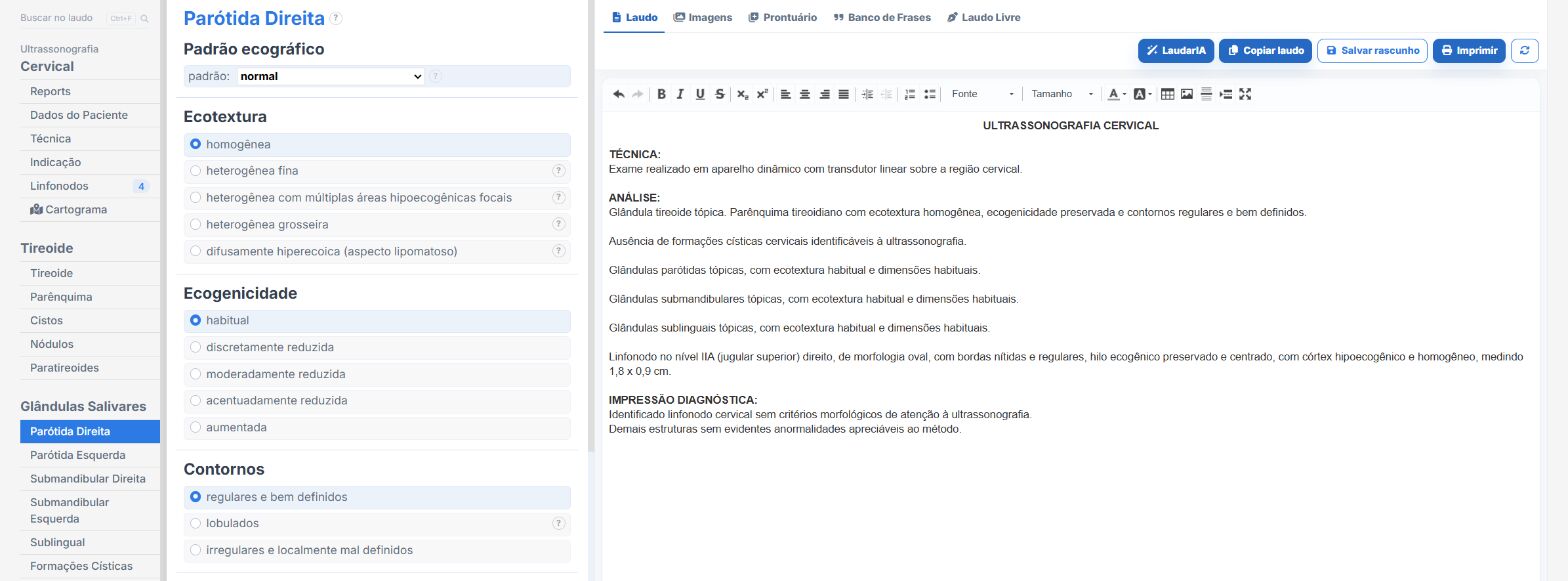
Laudário has a dedicated Salivary Glands template, with a tab for each parotid, each submandibular and the sublingual gland. In practice:
- Nodules with location by lobe, relative to the retromandibular vein, and by pole.
- Intraglandular or excretory duct stone, with ductal dilation.
- Acute and chronic sialadenitis, lipomatosis and the heterogeneous pattern with focal hypoechoic areas.
- Intraparotid and periglandular lymph nodes, with a cervical lymph node map.
- Recommendations in their own tab.
Try it free for 15 days, no credit card required.
The ultrasound reporting platform built by sonologists. We write about reports, classifications and standardization from the real routine of the practice. Who writes and how.
Educational content for healthcare professionals. It does not replace clinical judgment or the guidelines adopted by your service. Editorial policy.